lunedì 30 gennaio 2006

Post-traumatic fracture with cord compression


Findings

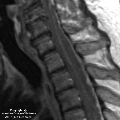
Two sagittal T2 images (Figure 1 and Figure 2) in a trauma patient. There is an acute compression fracture of the T11 vertebral body with retropulsion of the posterior cortex. There is loss of CSF signal between the posterior vertebral cortex and the spinal cord and there is deformity of the cord at the fracture site. Increased T2 signal is seen in the cord at this level.
Axial T1 image (Figure 3) demonstrates the fracture with associated cord compression, less well seen than on the T2 images. Abnormal signal adjacent to the cord is consistent with subacute hemorrhage.
Axial T2 image (Figure 4) confirms the fracture, loss of CSF between the cord and adjacent bone (Figure 4), and the two bright foci of signal abnormality within the cord. This increased T2 signal is seen within the central gray of the cord with sparing of the white matter tracts, a common initial presentation of cord edema. Additional bony disruption is seen on the left.


Diagnosis: Post-traumatic fracture with cord compression


Acute spinal cord compression is a potentially devastating neurological emergency that requires both prompt diagnosis and intervention to prevent permanent impairment. Close cooperation between clinical services and diagnostic radiologists is essential for patient triage. This is especially true in cases where patients cannot be fully examined neurologically. Magnetic resonance imaging is the study of choice in the evaluation of these patients, as it is noninvasive, does not involve radiation, and provides for investigation of both osseous and soft tissue lesions.

Information from the neurological exam is critical for localization of the lesion and optimization of the imaging protocol. Whole spine imaging is generally undesirable, as it is more time-consuming, expensive, and difficult for patients who are often in considerable pain. It further lowers resolution on exams that are often suboptimal secondary to severe patient pain and patient movement. Spinal sensory levels on neurological examination may be up to several segments below the anatomic level of cord compression. Evaluation of motor function and reflexes is very useful for lesion localization.

Once the site of interest is more precisely identified, sagittal T1 and T2 images and axial T2 images are required for the diagnosis. Axial T1 images through the lesion may then be obtained for further characterization of the anatomy and evaluation of hemorrhage. Intravenous contrast is not necessary for the diagnosis of acute cord compression.

Spinal cord compression may be defined as the presence of a mass lesion abutting the cord with the complete loss of intervening CSF. This must be accompanied by deformation of the spinal cord, or the presence of signal changes within the cord. The findings are best visualized on T2-weighted images. If the patient is concurrently symptomatic, acute intervention is mandated with the specific type of intervention determined by the underlying disease process. In acute cord compression secondary to trauma, imaging findings may also have prognostic value separate from findings on neurological exam.

martedì 24 gennaio 2006

Superior sagittal sinus thrombosis


Findings

Figure 1: Unenhanced head CT demonstrates prominent hyperdensity in the superior sagittal sinus. No parenchymal abnormalities are seen.
Figure 2: Sagittal T1 without contrast demonstrates abnormal hyperintensity within the superior sagittal sinus and the expected flow void is not seen.
Figure 3 and Figure 4: Contrast enhanced T1 images in the axial and coronal planes demonstrate abnormal hypointensity within the superior sagittal sinus surrounded by peripheral enhancement, the “empty delta” sign (Figure 3 and Figure 4). There is also uniform enhancement of the dural membranes.
Figure 5: Phase contrast MR venogram demonstrates no flow within the superior sagittal sinus. Flow is seen within the internal cerebral veins, the vein of Galen and the straight sinus.


Diagnosis: Superior sagittal sinus thrombosis


Superior sagittal sinus (SSS) thrombosis is an often underdiagnosed condition that can have serious neurologic sequelae. Thrombus occurring within the SSS and other intracranial venous structures may lead to cerebral venous infarction, hemorrhage, and hydrocephalus. Imaging characteristics of dural sinus thrombosis are key to its diagnosis as clinical symptoms are often variable and nonspecific. Common clinical signs and symptoms are headache, nausea, confusion, and lethargy.

Predisposing factors leading to SSS thrombosis may be grouped into one of three categories: hypercoagulable state, venous flow disturbance, and in association with infection or inflammation. However, up to one-quarter of cases are idiopathic. Hypercoagulable states may be congenital or acquired and include protein S deficiency, antithrombin III defeciency, oral contraceptive use, pregnancy, dehydration, and malignancy. Conditions in which there is disturbance of SSS flow include mass lesions and heart failure. Examples of infectious-inflammatory states predisposing to SSS thrombosis are sinusitis, mastoiditis, trauma, and sarcoidosis. The patient presented here was recently post partum, and a hypercoagulable state associated with her pregnancy was the most likely risk factor in the development of her SSS thrombosis.

On unenhanced CT, increased attenuation may be visualized within the SSS. The intracranial venous structures may appear prominent secondary to venous congestion. Contrast-enhanced CT and MR often demonstrate enhancement and/or enlargement of the venous structures. The “empty delta sign” may be present, which is likely caused by enhancing collateral channels in the dural membranes outlining a nonenhancing thrombus in the SSS. The sign is seen in only a minority of cases, but it is highly specific for SSS thrombosis. MR also may show lack of the expected flow void within the SSS, coupled with abnormal signal intensity within the SSS, suggesting the presence of thrombus. MR venography can show the lack of flow in the affected sinus. The MR venogram must be a phase contrast sequence as time-of-flight sequences can demonstrate hyperintense clot within the SSS that may be misinterpreted as patency. Secondary signs of the thrombus may also be present with T2- and diffusion-weighted abnormalities, venous congestion, hemorrhage, and hydrocephalus. SSS thrombosis is usually treated with anticoagulation. Most patients experience improvement, and many experience virtually complete recovery.


mercoledì 11 gennaio 2006

Spinal epidural abscess


Findings

Sagittal T1 (Figure 1): There is an isointense posterior epidural mass that extends inferiorly from the C4-C5 disc space level and displaces the spinal cord anteriorly.
Sagittal T2 (Figure 2): The posterior epidural high-signal mass is more well-defined on this T2-weighted image.
Axial T2 (Figure 3): Image demonstrates compression of cord elements by the posterior epidural mass.
Post-contrast Sagittal T1 (Figure 4): Image demonstrates an enhancing posterior epidural collection consistent with an abscess.


Diagnosis: Spinal epidural abscess


A spinal epidural abscess (SEA) can present with nonspecific signs and symptoms, including lower back pain, lower extremity weakness, or even sepsis. Cord compression can result if an epidural abscess in the spinal canal is not promptly treated. In a patient with back pain and fever, an SEA should be considered until proven otherwise.

An SEA can result through hematogenous spread or from direct extention of adjacent discitis or osteomyelitis. Remote infections, from indwelling catheters or even urinary tract infections, may hematogenously spread to create an SEA. Alternatively, any procedure in which there is direct puncture into the spinal canal, may seed an infection that leads to the development of an epidural abscess. Additional risk factors for the development of SEA include IV drug abuse, diabetes, alcoholism, and chronic immunosuppression.

MRI with gadolinium is the test of choice in evaluating patients suspected of having an epidural abscess. Alternatively, CT myelography can be utilized in patients who have contraindications to MRI. A lumbar puncture is a relative contraindication if a spinal epidural abscess is suspected, because infectious agents may be introduced into the subarachnoid space.

MRI findings usually fall into 2 categories: 1) A soft tissue mass that is hypointense on T1-weighted images and hyperintense on T2-weighted images with diffuse homogeneous or slightly heterogeneous enhancement within the collection; 2) As the phlegmonous mass necroses, there may be a peripherally-enhancing fluid collection. Spinal epidural abscesses may be located anteriorly or posteriorly within the spinal canal. Anterior epidural abscesses are generally secondary to spread of adjacent infection from discitis or osteomyelitis. Posterior epidural abscesses may occur as a result of hematogenous spread of remote infections. Other causes of back pain that should be considered in the differential diagnosis include herniated disc, neoplasm, spinal hematoma, and transverse myelitis.

Those patients who do not present with neurological symptoms may be treated with medical therapy alone. Those with neurological compromise are treated with surgical decompression and antibiotics. Our patient underwent successful surgical decompression of his spinal epidural abscess.

martedì 10 gennaio 2006

Medulloblastoma


Findings

Axial non-enhanced CT image demonstrates ill-defined slightly hyperdense lesion (Figure 1) centrally within the posterior fossa.
Axial non-enhanced CT image shows hydrocephalus with transependymal flow of CSF (Figure 2).
Axial T1-weighted image (Figure 3) demonstrates a posterior fossa mass that is hypointense on T1.
Axial T2-weighted image shows a well-defined heterogeneous mass (Figure 4).
Axial ADC map image (Figure 5) shows that the solid portions of the mass are dark, consistent with relatively decreased diffusion of water molecules.
Axial T1-weighted postcontrast image (Figure 6) shows a mass with heterogeneous contrast enhancement and areas of probable cystic change.
Sagittal T1-weighted postcontrast image demonstrates the large mass causing herniation of cerebellum and cerebellar tonsils inferiorly through the foramen magnum (Figure 7). The brainstem is compressed and shifted ventrally. There is compression of cerebral aqueduct that leads to marked hydrocephalus.

Differential diagnosis:
- Medulloblastoma
- Juvenile pilocytic astrocytoma
- Ependymoma
- Atypical teratoid
- Rhabdoid tumor


Diagnosis: Medulloblastoma


Medulloblastoma is thought to arise from undifferentiated neuroepithelial cells by neoplastic transformation of cells in the roof of the fourth ventricle and is categorized as a primitive neuroectodermal tumor (PNET). Medulloblastomas are the most common malignant central nervous system in children and the second most common pediatric brain neoplasm. It is one of the two most common primary tumors of the posterior fossa in children, the other one being juvenile pylocytic astrocytoma (JPA). Medulloblastoma is almost always found in the cerebellum, typically arising from the vermis. The tumor most frequently occurs in males under 10 years of age. Common presenting symptoms are headache, vomiting, and ataxia, and they are usually of less than 3 months in duration.

The classic CT appearance of medulloblastomas is a hyperdense, well-circumscribed, homogeneously-enhancing central cerebellar mass associated with obstructive hydrocephalus. On MR imaging, the tumors are typically of hypointense T1 signal and isointense to hypointense on T2-weighted images. There is a greater degree of heterogeneity among medulloblastomas on MR images compared to CT scans. Nearly all tumors show heterogeneous enhancement following gadolinium. The tumor appears heterogeneous due to hemorrhage, cystic change, and calcification, which occurs in 20% of cases. Medulloblastomas consist of small, densely packed cells (one of the "small blue cells" tumors), which leads to relatively decreased diffusion of water molecules seen as hypointensity on ADC maps. This feature appears to distinguish these tumors from JPA, which contain large interstitial spaces leading to increased diffusion and hyperintensity on ADC maps. Taurine peak may be detected within medulloblastomas on MR proton spectroscopy. Evidence of subarachnoid metastatic spread is present in up to one-third of cases at initial diagnosis.

Leptomeningeal involvement of the spinal cord is the most common site of spread and contrast-enhanced MRI is the imaging study of choice. Characteristic findings of spinal cord involvement are nodular enhancement of the cord surface or nerve roots, clumped nerve roots, and diffuse enhancement of the thecal sac. The use of ventriculoperitoneal shunts may lead to metastatic spread in the abdominal cavity. Tumor is very radiosensitive and thus, a combination of surgery and radiation treatment is most commonly used. Imaging of the entire brain and spinal cord is important to guide treatment prior to surgery. In addition to surgery and radiation, chemotherapy and shunt placement are also used in some cases, and postoperative chemotherapy without radiation has recently been found promising. A follow-up MRI with contrast is obtained postoperatively within 48 hours to assess for residual tumor before the development of enhancing reactive gliosis, which could be confused as tumor. Postoperative surveillance imaging of the brain and spine is regularly preformed, as recurrence is frequent. The majority of recurrences occur in the first two years after treatment. Recurrence presents as leptomeningeal enhancement or focal parenchymal nodular enhancement within the brain, most frequently in the posterior fossa, and metastatic involvement of vertebral bodies is sometimes seen.

mercoledì 4 gennaio 2006

Subclavian steal syndrome (SSS)


Findings

Doppler evaluation demonstrates reversal of flow in the left vertebral artery (Figure 1).
3D MIP reconstruction of a contrast-enhanced MRA of the aorta and great vessels demonstrates a focal left subclavian artery stenosis (Figure 2).


Diagnosis: Subclavian steal syndrome (SSS)


First described in 1961 by Reivich, et al, the triad of posterior fossa cerebral ischemia, unequal radial pulses, and stenosis/occlusion of the proximal subclavian artery was later dubbed the “subclavian steal syndrome” (SSS). Neurologic symptomatology indicates concomitant hemodynamically significant disease in the cerebral arterial circulation (such as inadequate Circle of Willis) or supply to this circulation (such as carotid stenoses). Only 5% of patients with evidence of angiographic steal, designated “subclavian steal phenomenon” (SSP), have associated neurologic symptoms.

Atherosclerotic disease is the primary underlying etiology of subclavian artery stenosis and SSS, accounting for the increased incidence of SSS in the elderly and in males vs females (1.5-2.1:1). Patients with SSS present with symptoms of vertebrobasilar insufficiency (dizziness, ataxia, and visual changes) and may also have arm claudication that is exacerbated with exercise or neck movements. Upon examination, there is diminished or absent radial/ulnar pulses, blood pressure reduction of greater than 20 mm Hg in the diseased arm or a proximal subclavian artery bruit.

Sonographic evaluation reveals decreased ipsilateral vertebral artery midsystolic velocity in mild subclavian artery stenosis and retrograde vertebral flow with associated tardus parvus or monophasic subclavian waveform in severe stenosis. Findings can be confirmed with MR angiography performed with phase-contrast, time-of-flight, or gadolinium-enhanced sequences. MRA demonstrates subclavian stenosis and reversal of vertebral flow.

Diagnostic and therapeutic angiography may be performed via a femoral, brachial or combined femoral/brachial approach. Subclavian stenoses may be treated with percutaneous transluminal angioplasty (PTA) or primary stenting. If PTA is utilized as the primary therapy, results should be confirmed with post-PTA hemodynamic evaluation. A suboptimal result can be improved with stent placement if intraarterial systolic pressure gradient across the lesion after PTA is =5 mm Hg or if there is =20% residual stenosis. Many operators choose to primarily stent subclavian artery stenoses. Three to 5 year patency rates of both PTA alone or combined PTA/stenting range from 86%-89%. Restenoses result from intimal hyperplasia. Most symptomatic restenoses occur within 26 months of intervention, and thus regular follow-up for at least 2 years post-PTA is recommended. Treatment of subclavian artery occlusion is also possible, although technical success is lower. Mortality is rare and minor complications are usually due to access site hematomas.

Surgical treatment options are less favored because of invasiveness and documented mortality rates of 0.4%-2.4%. These treatments include carotid-subclavian bypass with synthetic or vein graft, carotid-subclavian transposition or axilloaxillary bypass.

lunedì 2 gennaio 2006

Intradural lipoma


Findings

Sagittal T1, with and without fat supression, and sagittal T2-weighted sequences show an intradural hyperintense mass that supresses on fat saturation images (Figure 1, Figure 2, and Figure 3). Note chemical shift artifact on the T2-weighted sequence (Figure 3).


Diagnosis: Intradural lipoma


Intradural lipomas are the least common of the spinal lipomas, comprising 4%. Unlike this example case, they most commonly involve the cervical or thoracic cord, and are most commonly found dorsally, though they may lie laterally as well. Like other spinal lipomas, they are believed to be related to abnormal embryonic neurulation. When clinically symptomatic, intradural lipomas most commonly present with spinal cord compression.

Lipomyelomeningoceles, which account for 84% of spinal lipomas, can be thought of as similar to myelomeningoceles with associated lipomas, fibromuscular capsules, and intact overlying skin surface. Unlike myelomeningoceles, however, there is no association with Chiari II malformation. They may present with neurologic abnormalities, including neurogenic bladder, as well as associated osseous deformities.

Fibrolipomas of the filum terminale are of fat signal intensity, but are thinner and more linear in nature and may involve the filum itself and/or its lower dorsal dural attachment. Many are asymptomatic, although spinal lipomas as a group are the most common cause of cord tethering.