lunedì 29 giugno 2009

Lymphangioma of spinal canal


Findings


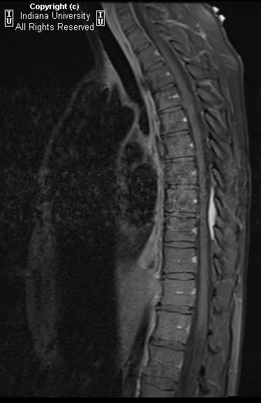
Homogeneously enhancing T1 isointense / T2 hyper intense well-defined extramedullary extradural lesion posterior to the mid-thoracic thecal sac. The lesion extends from the T8 superior endplate through T9 inferior endplate levels. Minimal posterior deformity the dorsal aspect of the thecal sac. No associated significant spinal canal stenosis or neuroforaminal compromise. No evidence of adjacent osseous invasion.

Differential diagnosis:
- Meningioma
- Metastasis
- Extramedullary hematopoiesis
- Lymphoma
- Lymphangioma


Diagnosis: Lymphangioma of spinal canal


Key points

Lymphatic malformation, benign hamartomatous, proliferation of lymph vessels.
Incidence estimated to be around 1 per 10 000 live births.
Fluid filled sacs that result from blockage of the lymphatic system.
Often associated with karyotype abnormalities such as Turner syndrome.
Usually divided into 2 major groups based on depth and size.
Superficial vesicles are called lymphangioma circumscriptum.
More deep-seated group includes cavernous lymphangioma and cystic hygroma, with many categorizing cystic hygroma as a variant of cavernous lymphangioma.
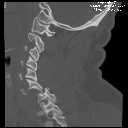
Most common sites are the head and the neck (75%), followed by the proximal extremities, the buttocks, and the trunk. However, they can be found anywhere, including the intestines, the pancreas, the mesentery, and the spine.
Typically seen in infants, (90%) are either evident at birth or become evident before age 2 years.
Rare in adults, the etiology in the adult population may be traumatic rather than congenital.

MR Findings:
- MRI can define the degree of involvement and the entire anatomy of the lesion.
- MRI can help prevent unnecessary extensive, incomplete surgical resection, because of the association with a high recurrence rate.

CT Findings:
- Low density, often poorly-circumscribed cystic mass, fluid-fluid levels may be seen in multi-loculated lesions.
- No significant enhancement in cystic uni- or multilocular neck mass.
- Wall is imperceptible & does not enhance if no infection.
- US can diagnose lymphangiomas, but MR or CT are usually necessary to map their entire extent.

Treatment:
- Surgical: preferred treatment is complete surgical excision if feasible. Image guided sclerosis with alcohol.
- Nonsurgical: No proven medical care for lymphangiomas exists, not responsive to radiation therapy or steroids.

martedì 23 giugno 2009

Ruptured spinal dermoid with chemical meningitis


Findings

Figure 1: Sagittal T1W image of the cervicodorsal spine shows multiple hyperintense foci in the subarachnoid space suggestive of fat deposition from dermoid rupture.
Figure 2 and Figure 3: Sagittal T1W and T2W images reveal a heterogeneous hyperintense mass expanding the lower spinal cord and conus medullaris.
Figure 4 and Figure 4: Axial T1W images demonstrate hyperintense signal within the central canal.
Figure 6 and Figure 7: Axial T1W images of brain show fat droplets, seen as hyperintense foci in the peri-mesencephalic cistern and in the frontal horn of left lateral ventricle.

Differential diagnosis
- Spinal dermoid
- Teratomas
- Lipomas

Differential diagnosis of brain lesions that appear hyperintense on T1W images on brain MRI are as follows:
- Lesions with hemorrhagic components (such as infarct, encephalitis, intraparenchymal hematoma, cortical contusion, diffuse axonal injury, subarachnoid hemorrhage, subdural and epidural hematoma, intraventricular hemorrhage, vascular malformation and aneurysm, and hemorrhagic neoplasm)
- Protein-containing lesions (such as colloid cysts, craniopharyngiomas, Rathke's cleft cysts and atypical epidermoids)
- Lesions with fatty components (such as lipoma, dermoid and lipomatous meningioma)
- Melanin-containing lesions (such as metastasis from melanoma and leptomeningeal melanosis)
- Lesions with calcification or ossification (such as endocrine-metabolic disorder, calcified neoplasm, infection, and dural osteoma)
- Lesions with other mineral accumulation (such as acquired hepatocerebral degeneration and Wilson disease)
- Miscellaneous group (which includes ectopic neurohypophysis, chronic stages of multiple sclerosis and neurofibromatosis type I)


Diagnosis: Ruptured spinal dermoid with chemical meningitis


A spinal dermoid is a rare, benign, slow-growing, dysontogenetic tumor arising from the inclusion of ectopic embryonic rests of the ectoderm and mesoderm within the spinal canal at the time of neural tube closure between the 3rd and the 5th week of fetal life. Several causes including spontaneous, iatrogenic or traumatic rupture have been reported to result in dissemination of lipid material from the dermoid tumours into the subarachnoid space or ventricles. Intra-spinal dermoids account for approximately 1–2% of intra-spinal tumors and tend to extend to the subarchnoid space, however there is no communication between the cyst and the subarchnoid space. They occur predominantly in the lumbosacral region and rarely in the cervicothoracic region. They show a slight male predominance, and most reveal themselves during the second and third decades.

Pathologically, spinal dermoids have a thick wall covered with stratified squamous epithelium containing dermal appendages such as hair, sebaceous glands and sweat glands and less commonly, teeth and nails. Dermoid tumours commonly have areas of calcification. Their rupture and spread is a rare event, which may be asymptomatic or may present acutely as chemical meningitis. The exact incidence of rupture of spinal dermoids is not known. Cholesterol, which is the most irritating element of the dermoid excites an inflammatory/ granulomatous meningeal reaction.

Spinal dermoids appear as variegated, space-occupying lesions showing a heterogeneous signal on T2-weighted MRI due to the different components within the cyst. These have high protein-containing fluid and thereby appear brighter than CSF on T1-weighted images. The high lipid content also gives a characteristic hyperintense signal on T1-weighted spin-echo images.

In conclusion, radiologists need to be aware that whenever a diagnosis of intra-spinal dermoid is suggested, it is essential to screen the entire neural axis to look for possible rupture and CSF spread. MRI is the imaging modality of choice.

venerdì 19 giugno 2009

Lymphocytic hypophysitis


Findings

Figure 1: Thickened pituitary infundibulum.
Figure 2 and Figure 3: Enhancing thickened pituitary infundibulum.


Diagnosis: Lymphocytic hypophysitis


Lymphocytic hypophysitis is an inflammatory lesion of the pituitary gland characterized by lymphocytic infiltration. The disease was first recognized in 1962 by Goudie and Pinkerton. The disease is rare, with a striking female predominance (approximately 9:1). As of 1997, only six cases had been diagnosed in men; however, definitive diagnosis requires pathologic correlation either through surgery or autopsy. There is a predilection for the disease during pregnancy and the postpartum period.

Radiologically, the disease is characterized by homogeneously enhancing pituitary mass with infundibular thickening. This is often difficult to distinguish from an atypically enhancing macroadenoma; however, the finding of dural enhancement or extrapituitary involvement within the subarachnoid space can favor the diagnosis of lymphocytic hypophysitis.

Clinically patients present with either partial/total hypopituitarism or symptoms related to mass effect. Treatment consists of corticosteroids with replacement of any deficient hormones.

giovedì 18 giugno 2009

Os Odontoideum


Findings

The coronal CT neck image in Figure 1 shows a small, round os odontoideum separated from the body of C2 by a wide radiolucent gap. It has smooth and uniform cortical margins.
The sagittal CT neck image in Figure 2 shows signs of C1 anterior arch hypertrophy. There is no paravertebral swelling. The posterior atlanto-dens interval (PADI) also known as the space available for cord (SAC) is 12mm (less than 13mm has a poor prognosis).


Diagnosis: Os Odontoideum


Os odontoideum describes a rare condition in which the dens is separated from the axis body. Two types of os odontoideum have been identified. An orthotropic os odontoideum is located in the normal position of the odontoid process, whereas a dystrophic os odontoideum is either attached to the anterior arch of C1 or the clivus.

The etiology of os odontoideum is still debated. The leading hypothesis suggests that trauma in early childhood causes injury to the soft tissues between C1 and C2. This is often seen when babies fall from their cribs or toddlers fall from stairs. Radiographic images taken after this injury usually do not show any significant pathology besides retropharyngeal swelling.

Injury results in avascular necrosis of the odontoid process over a period of months or years. Therefore, the cephalad ossification centers do not fuse with the C2 body. With growth, the alar ligaments that attach the tip of the dens to the occiput pull the ossicle upwards giving it its characteristic appearance.

Os odontoideum may also occur in patients with congenital anomalies including Down's syndrome, multiple epiphyseal dysplasia, Klippel-Feil malformation and Morquio syndrome. These patients are born with deficient odontoid processes.

Symptoms are variable with vague pain in the neck and shoulders being the most common. Less commonly reported symptoms include headaches, torticollis, weakness and paresthesias. The most serious complications of an os odontoideum occur when C1-C2 instability causes spinal cord compression or vertebral artery compromise. This may result in brain stem symptoms, quadraparesis, transient bouts of unconsciousness and sudden death.

The best way to evaluate an os odontoideum is by obtaining open-mouth anterior-posterior and flexion-extension lateral plain films. A criteria often used to make the diagnosis is the Posterior AtlantoDens Interval (PADI) also known as the Space Available for Cord (SAC). This is the distance between the posterior border of the dens and the anterior border of the posterior ring of the atlas. A PADI of less than 13mm is associated with neurologic decline. C1-C2 translation of more than 5mm on flexion-extension radiographs also has a poor prognosis.

The most common treatment for a symptomatic os odontoideum is C1-C2 posterior arthrodesis. This is particularly indicated in patients who exhibit signs of cord compression. Medical management is indicated for patients with only mechanical symptoms.

venerdì 12 giugno 2009

Osseous Epithelioid Hemangioendothelioma


Findings

On the non-contrast thoracic spine CT (Figure 1), there is a lytic lesion within the T5 vertebral body, extending into the posterior elements (red arrrow in Figure 2). The lesion is associated with an adjacent soft tissue mass (yellow arrow in Figure 3). The lytic areas have thin sclerotic margins, resulting in a distinctive “soap-bubble” appearance (yellow arrow in Figure 4). There is mild bony expansion of the T5 vertebral body, causing moderate spinal canal stenosis. There is no internal matrix formation, nor is there evidence of periostitis.


Diagnosis: Osseous Epithelioid Hemangioendothelioma


An epithelioid hemangioendothelioma (EH) is a rare intermediate-grade neoplasm of the vascular endothelial cells. EH can occur in bones, skin or viscera (liver, lung or spleen). The most common bones to be affected are the tibia (23%), femur (18%), humerus (13%) and vertebral bodies (10%). Rarely, the skull or pelvis may be involved. Osseous EH often demonstrates a regional pattern of involvement and there is frequently more than one lesion found within a single extremity. Osseous EH most often occurs in young patients, typically in the second or third decades of life. Males are affected twice as often as females. Patients usually present with pain and pathologic fractures are present in 10% of cases. If the tumor is located within the spine, the patient may experience neurologic symptoms. Rarely, patients will present with hemolytic anemia or a consumptive coagulopathy.

On radiographs and non-contrast CT, an osseous EH is an osteolytic lesion without matrix mineralization. Osseous EH typically has a lytic bubbly appearance often associated with expansile bony remodeling. There may be cortical break-through or surrounding sclerosis. A periosteal reaction is notably absent unless there is an associated pathologic fracture. An adjacent soft tissue mass may or may not be present. Lesions with ill-defined margins and loss of trabeculae are usually more aggressive in nature than well-circumscribed tumors. Osseous EH most frequently occurs in the tubular bones of the lower extremity and is often multifocal within the extremity. Joint invasion is common. In the spine osseous EH has a distinctive “soap-bubble” appearance with sclerotic margins. When osseous EH occurs in the spine, osteolysis may be present within adjacent vertebral bodies with narrowing of the intervening disc space mimicking osteomyelitis/discitis. If intravenous contrast is administered, EH demonstrates marked homogenous enhancement which is to be expected since it is a vascular tumor. On MRI, EH is generally T1 hypointense, T2 hyperintense, and markedly hypervascular. Osseous EH results in focal uptake on bone scan. Multiple modalities are often needed to assess for multifocal bone or visceral involvement including bone scan, skeletal survey, and CT.

Differential considerations include metastatic disease (particularly melanoma, lung, breast, renal and thyroid cancer), angiosarcoma (indistinguishable), hemangiopericytoma, osteomyelitis/discitis, Ewing sarcoma, skeletal angiomatosis, telangiectatic osteosarcoma, primary lymphoma of bone, aneurysmal bone cyst, and Langerhans cell histiocytosis.

Osseous EH has a wide spectrum of behavior, but is usually locally aggressive. There is a 31% rate of metastatic disease, usually to the lungs or bone. Treatment is surgical excision, either curettage or wide resection, depending on the location and tumor grade. Adjuvant radiation therapy may be employed for aggressive tumors. The role of chemotherapy is controversial. As expected, well-differentiated tumors have a favorable prognosis following resection but high grade tumors have a dismal prognosis. Approximately 13% of tumors will have a local recurrence. The most important predictor for prognostic purposes is associated visceral involvement the presence of which indicates a poor prognosis.

martedì 9 giugno 2009

Paradichlorobenzene intoxication with encephalopathy


Findings

Shown are proton density and T2 axial images of the patient’s brain. The white matter is diffusely hyperintense on long TR sequences, including corticospinal tracts in the brainstem, cerebellar white matter and corpus callosum. The thalami and basal ganglia appear diffusely hypointense on long TR sequences.


Diagnosis: Paradichlorobenzene intoxication with encephalopathy (recreational mothball sniffing and eating)


Paradichlorobenzene (p-DCB, PDB; 1,4-DCB) is a volatile chlorinated hydrocarbon, a fat-soluble chemical. The primary exposure to 1,4-dichlorobenzene is from breathing contaminated indoor air. Acute (short-term) exposure to 1,4-dichlorobenzene, via inhalation in humans, results in irritation of the skin, throat, and eyes. Chronic (long-term) 1,4-dichlorobenzene inhalation exposure in humans results in effects on the liver, skin, and central nervous system (CNS). No information is available on the reproductive, developmental, or carcinogenic effects of 1,4-dichlorobenzene in humans.

Paradichlorbenzene (which constitutes over 95% of the content of mothballs), may be used as a recreational drug. There are very few cases reported of mothball abuse and toxicity, mostly due to underreporting of this habit by the patients, who are usually teenagers and young adults. Our patient, a 29 year old woman, presented to the hospital with complaint of generalized malaise, progressive mental decline over several weeks and headache leading to evaluation with brain MRI. The patient’s mental status had progressively deteriorated with no focal abnormalities on neurological exam. 6 days into her hospital stay, the patient became obtunded, responding to her name sluggishly and was unable to speak coherently. The woman’s laboratory studies revealed transaminitis and iron deficiency anemia. Note was made of faint smell of mothballs present in the patient’s room since her hospitalization. On focused questioning, the family reported an an odor of mothballs in her apartment for months. The patient explained this away as “getting ready for spring and cleaning out her closets.” After these facts were put together, the blood level of paradichlorbenzene was obtained and measured 15 mcg/mL, the highest level ever recorded in medicine, with normal being undetectable. The history emerged of her inhaling and EATING mothballs for months as a recreational mind-altering activity.

Paradichlorbenzene is a fat-soluble chemical and accumulates within body fat. The fat can serve as a long term repository. In high concentrations it may perpetuate patient’s clinical deterioration for a long time after cessation of substance abuse.

Differential diagnosis for MRI findings of paradichlorbenzene intoxication include toluene toxicity and carbon monoxide poisoning. In toluene intoxication, T2 fast-spin echo sequence shows diffuse white matter hyperintensity with hypointensity of thalami, basal ganglia, red nuclei and pars reticulata of the substantia nigra. In CO poisoning, T2 and FLAIR sequences show high signal within the globus pallidi, which are most sensitive to carbon monoxide poisoning, followed by involvement of the caudate nucleus, putamen and thalamus. Involvement of the brainstem and cerebellum may be a reflection of more severe poisoning, but the cerebral white matter is not involved.

venerdì 5 giugno 2009

Bilateral jumped facets with spinal cord transection


Findings

Axial CT scan of the cervical spine demonstrates the naked facet sign, with uncovered articulating processes. A fracture is noted anteriorly on the left.
Sagittal image shows marked subluxation of C6 on C7 with marked narrowing of the spinal canal.
Sagittal images of the right facets. The facets are jumped.
Sagittal T2 and STIR show marked anterior subluxation of C6 on C7, transection of the spinal cord, edema and hemorrhage in the prevertebral soft tissues and spinal cord edema extending from C4 to T1.


Diagnosis: Bilateral jumped facets with spinal cord transection


Bilateral facet joint dislocation is a severe unstable cervical flexion injury. Flexion- rotation injuries can cause dislocation of the vertebral bodies with jumped/locked facets which can be unilateral or bilateral. If the dislocation is not complete, the facets may appear “perched” upon one another. Unilateral facet dislocation more characteristically has a rotatory component and carries a better prognosis.

With complete dislocation, the facets are said to be jumped and/or locked. The integrity of all the spinal ligaments including the posterior longitudinal ligament, the posterior portions of the annulus fibrosus, and the capsular, interspinous and supraspinous ligaments is disrupted. Destabilization of the ligamentous support of the cervical spine allows for the charactersitic >50% anterolisthesis observed. Unfortunately, severe narrowing of the spinal canal and intervertebral foramen occurs with subsequent severe neurologic deficits (in approximately 75% of cases), as seen in this case with spinal cord transection.

The naked facet sign, also referred to as the, “Hamburger Sign” refers to the CT appearance of uncovered articulating processes. Normally, on axial CT, the vertebral facet joint looks like a hamburger with the superior articular process of the vertebra below forming the, “bun” on top of the “meat patty,” and the inferior articular process of the vertebra above forms the bun beneath the patty. When the joint is dislocated, the superior articular facet (top bun of the Hamburger) now lies posteriorly. This is the Hamburger Sign, which can be unilateral or bilateral.

Associated osseous injuries include avulsion of the spinous process above the locked level and small triangular fractures of the anterosuperior corner of the inferior involved vertebral body.

giovedì 4 giugno 2009

Anterior subluxation of C4 on C5, with other imaging demonstrating unilateral locked left facet and perched right facet at C4-C5.


Findings

Figure 1: Lateral Cervical spine plain film demonstrates anterior subluxation of C4 on C5 and widening of spinous processes.
Figure 2: Sagittal MRI image shows anterolisthesis of C4 on C5 with central disc herniation. The spinal cord demonstrates normal signal intensity. Increased T2 signal intensity is present posteriorly, consistent with ligamentous disruption with edema and hematoma of posterior soft tissues.


Diagnosis: Anterior subluxation of C4 on C5, with other imaging demonstrating unilateral locked left facet and perched right facet at C4-C5.


Hyperflexion injuries can occur during trauma and are described as a combination of distraction and flexion that disrupts the ligamentous structures between two adjacent vertebrae, but the anterior longitudinal ligament remains intact. The height of the anterior interspace is narrowed and the posterior height is widened. There is also a possibility of disruption of the facet joints. Hyperflexion can result in injury to the posterior and middle columns whereas hyperextension may lead to injury to the anterior and/or middle columns.

A unilateral locked facet is secondary to excessive flexion and rotation and is considered a stable injury. The inferior facet of the superior vertebra moves up and anterior to the inferior vertebra thereby causing them to sit in a “locked” position. In unilateral locked facets there is typically anterolisthesis of the superior vertebral body of <50% of the width of the vertebral body. If there is translation of more than 50%, then there is most likely bilateral locked facets, which is an unstable injury.

Cervical spine CT with sagittal reconstructions or lateral cervical plain films can be used for measuring cervical translation. Lines are drawn along the involved posterior vertebral bodies and the transverse distance is measured between the lines at the level of the inferior endplate of the upper vertebra.

MRI may be used in patients with acute cervical spine trauma to evaluate neurologic or ligamentous integrity. It is indicated in patients with negative radiographs and negative CT who have neurologic symptoms, and also in patients with fracture or unstable injury. It has recently been noted that MR imaging has high sensitivity for most ligamentous injuries, in particular posterior longitudinal ligament, interspinous ligaments, disc annulus and facet capsular ligaments. Sensitivity is somewhat lower for anterior longitudinal ligament and ligamentum flavum injury after comparing with intraoperative findings. It should also be noted that, while sensitive, MR findings were generally not found to be highly specific with respect to intraoperative findings and can result in overestimation of ligamentous injury.

lunedì 1 giugno 2009

Creutzfeldt-Jakob Disease (CJD)


Findings

Noncontrasted head CT shows subtle basal ganglia hypo attenuation bilaterally and enlarged bilateral extra-axial fluid spaces. Contrast enhanced brain MR demonstrates increased signal on FLAIR and T2-weighted sequences with corresponding restricted diffusion bilaterally in the caudate nuclei, globi pallidi, posterior thalami, hippocampi and in the cortex of the bilateral occipital-parietal, right frontal and temporal lobes without enhancement. Brain MR 4 weeks later shows persistence of restricted diffusion in the same areas of the brain.

Differential diagnosis:
- Mitochondrial disorder like MELAS or Leigh syndrome
- Creutzfeldt-Jakob Disease
- Viral encephalitis
- Carbon monoxide poisoning


Diagnosis: Creutzfeldt-Jakob disease.


Key points

CJD is one of the neurodegenerative disorders called prion diseases (also known as spongiform encephalopathies), which are uniformly fatal.
90% of cases are sporadic; the other 10% are familial.
CJD is characterized clinically by rapid and progressive dementia, other neurologic abnormalities, and ultimately death.
There is no cure.
WHO criteria for diagnosis requires tissue for definite cases or typical clinical, lab or EEG findings for probable or possible cases; imaging findings are not included.
Typical MR findings of sporadic CJD include increased signal in grey matter on T2-weighted, FLAIR, proton density, and diffusion-weighted (DW) images.
It is more often bilateral.
The caudate nuclei and putamina are usually more affected than the thalami and cerebral cortex.
Diffusion-weighted images are the most sensitive sequence for early, typical signal changes, followed by FLAIR sequences.
Eventually, brain atrophy is a characteristic feature of CJD.
Persistent restricted diffusion, over weeks or months, in typical areas of the brain is virtually diagnostic of CJD.
Acute diffusion restriction in the basal ganglia is seen with carbon monoxide poisoning, but it does not persist and preferentially involves the globus pallidus.