Visualizzazione post con etichetta Pediatric. Mostra tutti i post

lunedì 14 marzo 2011

Band heterotopia

CASE 1 (7-month-old)


CASE 2 (Pre-adolescent)


Findings

Case 1: Axial and coronal T2-weighted images of the brain in a 7-month-old girl with seizures demonstrate a band of isointense signal within the subcortical white matter, characteristic of band heterotopia.
Case 2: Axial and coronal T1-weighted images demonstrate band heterotopia, better seen in this preadolescent girl due to completion of myelination.


Diagnosis: Band heterotopia


Band heterotopia is a rare neuronal migration anomaly which manifests as homogenous bands of gray matter are interposed between the lateral ventricles and cortical mantle with normal appearing white matter on either side. The overlying cortex may be normal, pachygyric, or display a simplified gyral pattern with short gyri and shallow sulci. At least six morphologically distinct subtypes have been described. Band heterotopias represent a subset of gray matter heterotopia which also includes subependymal and subcortical heterotopia subtypes.

Band heterotopia typically affects female patients as a result of an X-linked dominant inheritance pattern secondary to abnormal function of the doublecortin (DCX) gene (Xp22.3-p23) or less frequently the LIS1 (17p13.3) gene. Male patients can be affected due to sporadic mutations of these genes (41 reported cases in the literature by D’Agostino, et al in 2002). The rate of detectable mutations involving DCX or LIS1 in male patients (42%) is lower than the rate of 85% described in female patients. Dysmorphic features described in patients with band heterotopia include microcephaly (most common), wide nasal bridge, high arched palate, and short stature.

The clinical presentation of band heterotopia can range from normal to nearly normal intelligence and mild developmental delay to frank mental retardation. Seizures are often also present and may begin in the first decade, ranging from partial to generalized or multiple seizure types. The discovery of the underlying brain malformation is due to the onset of seizures in 65% of patients. Eventually 95% of patients with band heterotopias will develop epilepsy. Seizures associated with band heterotopia are often refractory to medical therapy, and surgical therapies such as callosotomy may be performed in these patients. In the series of 30 male patients published in 2002, 46% of patients were refractory to medical therapy and experienced up to 20-30 seizures daily despite trails of multiple therapeutic regimens. Affected male patients tend to have either mild or severe symptoms, whereas, female patients tend to have symptoms within the mild to moderate range of the spectrum from minimal cognitive impairment to severe mental retardation. Posterior involvement, in particular the partial posterior and intermediate posterior subtypes, occur more commonly in male patients. Frontal and diffuse subtypes are more often present in affected female patients.

venerdì 10 dicembre 2010

Capillary Telangiectasia


Findings

There is an ill-defined enhancing focus in the medial right temporal lobe on post gadolinium contrast T1-weighted imaging (Figure 4). There is no corresponding signal abnormality or mass on the precontrast T1-weighted, T2-weighted, or FLAIR images (Figure 1, Figure 2, and Figure 3, respectively). There is no mass effect. On susceptibility-weighted imaging (SWI) the lesion shows hypointensity (Figure 5).


Diagnosis: Capillary Telangiectasia


Brain capillary telangiectasias are benign vascular malformations which are often found incidentally.
They can be visualized by gadolinium contrast and gradient-echo susceptibility or susceptibility weighted imaging, but not through catheter angiography, and may often not be visible on conventional T1/T2, FLAIR, or diffusion-weighted imaging.
Often asymptomatic and usually no treatment is required.

Brain capillary telangiectasias (BCTs) are one of four major types of vascular malformations which occur in the brain (the other three are arteriovenous malformations, cavernous malformations (cavernous angiomas), and developmental venous anomalies (venous angiomas), and represent up to 20% of all intracranial vascular lesions. BCTs consist of multiple ectatic capillaries surrounded by normal brain parenchyma and are usually devoid of calcification, gliosis, extraluminal hemorrhage, and hemosiderin-laden macrophages. BCTs are most common in the midbrain, pons, medulla, and spinal cord, but they are found throughout the central nervous system. Multiple BCTs are possible, especially in certain syndromes (e.g.; ataxia telangiectasia, Osler-Weber-Rendu, or Sturge-Weber syndrome).

Often found incidentally, BCTs are usually benign, small in size, and rarely grow over time. They are rarely symptomatic and are not associated with any particular clinical feature but have been reported to be associated with headache, vertigo, and tinnitus.

BCTs are relatively well visualized through susceptibility weighted imaging where they demonstrate marked signal intensity loss due to deoxyhemoglobin present in slow flowing blood. They are also well visualized through gadolinium-enhanced T1-weighted imaging sequences where they are seen as small faint lesions. BCTs are difficult to visualize through conventional T1/T2, FLAIR, or diffusion-weighted imaging and are considered to be one of the “angiographically occult vascular malformations” due to their small size, tendency to occlude, and sluggish flow.

venerdì 26 novembre 2010

Ewing sarcoma of the occipital bone


Findings

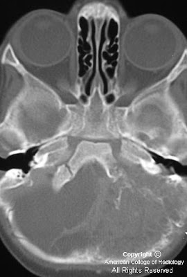
Figure 1: Unenhanced CT shows a heterogeneous attenuation mass with cystic spaces in the posterior fossa.
Figure 2: Intense enhancement is noted after contrast administration.
Figure 3: Bone window section showing permeative destruction of the left occipital bone.
Figure 4: Axial T1 weighted image showing extra- axial mass with multiple cystic spaces.
Figure 5: Coronal T2 image showing the extra-axial origin clearly with mass effect on the cerebellum. The cystic spaces appear hyperintense.
Figure 6: Axial T1 weighted image showing intense enhancement of the mass.


Diagnosis: Ewing sarcoma of the occipital bone


Ewing sarcoma is a small round-cell tumor arising from mesenchymal cells. These tumors affect children and young adults in the age group of 5-15 years. The long bones, flat bones like the scapula and the vertebrae are the most common sites. Primary Ewing sarcoma affecting the calvarium is extremely rare, making just 1% of the cases. In the skull, the tumor more often arises from the frontal and parietal bones and less common locations include ethmoid, temporal and occipital bones.

CT scans (bone window) reveal poorly marginated permeative destructive lesion involving both inner and outer tables of the skull. The "onion peel" appearance typical of Ewing sarcoma in long bones is not seen commonly in the calvarium. The extra-dural soft tissue shows intense enhancement on contrast administration.

MR imaging provides better soft tissue delineation of these tumors. The extra dural soft tissue appears hypointense on T1 weighted images while the cystic and necrotic areas appear hyperintense on T2 weighted images. Good contrast enhancement is noted.
The differential diagnosis should include rhabdomyosarcoma, metastatic neuroblastoma and lymphomas.

Treatment is surgery followed by chemotherapy and radiation.

lunedì 22 novembre 2010

Suprasellar arachnoid cyst


Findings:

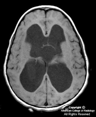
Figure 1, Figure 2 and Figure 3 show severe hydrocephalus involving the lateral and third ventricles.
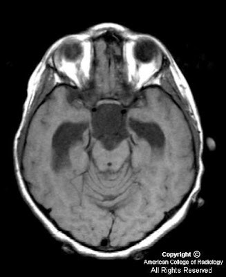
Figure 4, Figure 5, Figure 6, show a suprasellar mass lesion which follows CSF signal on all sequences, including FLAIR.


Diagnosis: Suprasellar arachnoid cyst


Arachnoid cyst is the most common congenital lesion of the brain. It is typically an incidental imaging finding and is rarely symptomatic. However, in this case the size and location of the lesion in the suprasellar cistern resulted in severe obstructive hydrocephalus and precocious puberty. Both of these complications resolved following surgical drainage of the cyst.

Arachnoid cysts arise from a splitting of the arachnoid membrane with formation of a cyst wall consisting of fibrous connective tissue. There is no epithelial lining in the wall. Expansion occurs following trapping of cerebral spinal fluid through defects in the cyst wall. Arachnoid cysts occur most commonly in the middle cranial fossa and have a 4-to-1 male-to-female ratio. Even large cysts tend to be asymptomatic. Associated clinical features in symptomatic patients include headache, calvarial bulging, intracranial hypertension, craniomegaly, developmental delay, visual loss, precocious puberty, and seizures. Treatment of arachnoid cysts is not recommended by many unless there is a clear cause and effect relationship between the cyst and symptoms as shown in this case.

MRI is the preferred diagnostic modality for arachnoid cysts because of its ability to demonstrate the location, extent and relationship of the cyst to surrounding neurologic structures. Lesions typically have the signal intensity of CSF on all sequences, do not enhance and do not demonstrate restricted diffusion. The most important differential diagnostic consideration is between arachnoid and epidermoid cysts. Epidermoid cysts show restricted diffusion on diffusion-weighted images. In addition, unlike epidermoid cysts, arachnoid cysts show suppressed signal on fluid-attenuated inversion recovery (FLAIR) images.

lunedì 8 novembre 2010

Neuroblastoma metastases


Findings

Figure 1: Axial post gadolinium T1 weighted image showing solid enhancing parenchymal lesion in the right temporal lobe with dural and leptomeningeal disease.
Figure 2: Coronal post gadolinium image showing dural enhancement along the tentorium and leptomeningeal enhancement.
Figure 3: Axial susceptibility weighted imaging revealing the hemorrhagic nature of the lesion.


Diagnosis: Neuroblastoma metastases


Neuroblastoma metastatic to the central nervous system is extremely rare, and the reported incidence varies from 1% to 16% at recurrence. Paediatric tumors that metastasise to the brain, in order of frequency, include neuroblastoma, osteosarcoma, Ewing sarcoma, rhabdomyosarcoma and Wilm tumor.

Risk factors for developing intracranial metastases include lumbar puncture at diagnosis, ages 2 to 3 years, bone marrow involvement, and MYCN gene amplification. Newer chemotherapeutic agents with better activity fail to penetrate the blood-brain barrier, thus facilitating a sanctuary for tumor cells within the central nervous system. As a result, the metastases evolve and become extensive before becoming clinically evident. Metastatic spread of tumor cells to central nervous system may occur either via hematogenous or cerebrospinal fluid routes and involve the neuroparenchyma, leptomeninges or dura.

Neuroparenchymal metastases from neuroblastoma have varied appearances. They may be cystic lesions with calcified mural nodules. The wall and mural nodules show intense enhancement with contrast. Metastases may also be solid and hemorrhagic and show homogeneous enhancement. Gradient or susceptibility weighted imaging would help in detecting hemorrhagic components. Leptomeningeal and dural metastatic involvement if present indicates poor prognosis.
lunedì 1 novembre 2010

Retinoblastoma with recurrence following enucleation


Findings

Figure 1: Contrast enhanced CT scan shows a heterogeneously enhancing intra-ocular mass in the right eye with calcification.
Figure 2: Contrast enhanced CT image shows enlargement of the right optic nerve suggestive of orbital recurrence following enucleation. Note the presence of a prosthetic globe.


Diagnosis: Retinoblastoma with recurrence following enucleation


The differential diagnosis of leukocoria in a child includes retinoblastoma, Coat’s disease, toxocariasis, retinopathy of prematurity, optic nerve drusen, persistent hyperplastic primary vitreous and phthisis bulbi. With a normal sized globe and intra-ocular calcifications the differential narrows down to retinoblastoma, Coat’s disease and toxocariasis with retinoblastoma being the most common.

Most patients with unilateral, non-metastatic retinoblastoma can be cured with enucleation alone. In patients with histologic risk factors, adjuvant chemotherapy is recommended, with the addition of orbital radiation for patients with trans-scleral involvement or tumor present at the level of the cut end of the optic nerve. Patients with metastases require intensive chemotherapy and consolidation with autologous hematopoietic stem cell rescue.

Recurrent retinoblastoma or a second primary neoplasm may be observed during follow-up. Optic nerve invasion, scleral invasion and choriodal invasion have been regarded as risk factors for recurrent disease. In general recurrent tumors have a short latent period as compared to second primary neoplasms; however this may not always be true. Recurrent retinoblastoma after enucleation can occur in the contralateral eye (type A), in the ipsilateral orbit (type B) or extend from the orbit into the brain (type C)

The most common presentation of orbital recurrence is a clinical complaint such as eyelid swelling or chemosis, suggesting that clinicians should not attribute periocular or orbital symptoms during the first year after enucleation as a sign of a benign conjunctivitis or preseptal cellulitis. A problem with the ocular prosthesis is also a frequent mode of presentation in these patients, and extrusion or displacement of a previously satisfactory prosthesis should be considered a suspicious sign for possible tumour recurrence.

Calcification maybe seen within the recurrent tumors on unenhanced CT. Most recurrent tumors appear iso to hypointense to normal temporal muscle on T1 weighted images and show moderately high signal on T2 weighted images. Contrast enhanced scans, CT or MRI, show heterogeneous enhancement of the tumor.

Cases with recurrence following enucleation should be evaluated for distant metastasis. Treatment consists of systemic chemotherapy combined with other modalities like orbital radiotherapy and stem cell rescue, depending upon the extent of metastatic disease.

venerdì 29 ottobre 2010

Pott's Puffy Tumor


Findings

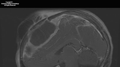
Figure 1: The image shows a large collection anterior to the frontal bone and a large epidural abscess with peripheral enhancement.
Figure 2: Erosion of the frontal bone. Sinus tract of the bone into the large anterior collection.
Figure 3: Large collection anterior to the frontal bone.
Figure 4: Large epidural abscess.
Figure 5: Large epidural abscess with peripheral enhancement.
Figure 6: Large collection anterior to the frontal bone. Large epidural abscess. Normal bone marrow signal. Abnormal bone marrow signal in the frontal bone.
Figure 7: Large collection anterior to the frontal bone with peripheral enhancement. Large epidural abscess with peripheral enhancement.
Figure 8: Abnormal bone marrow signal in the frontal bone. There is a lack of enhancement of the frontal bone. The abnormal dark bone marrow signal on T1 images and lack of enhancement on T1 post gad fat sat is consistent with dead necrotic bone from severe osteomyelitis.


Diagnosis: Pott's Puffy Tumor


Pott’s puffy tumor is a subperiosteal abscess of the frontal bone that appears as a localized swelling of the forehead associated with frontal osteomyelitis. Pott’s puffy tumor is a complication of frontal sinusitis or trauma, which is predominatly seen in the adolescent age group. However, there are a few case reports in adults. Pott’s puffy tumor is a rare complication of frontal sinusitis in the post antibiotic era but can be seen in patients with undiagnosed or partially treated sinusitis. Patients will typically present with frontal scalp swelling, headache, fever, nasal drainage, and frontal sinus tenderness. Ocassionally, Pott’s puffy tumor can mimic findings of preseptal or orbital cellulitis. In severe cases, there will be neurologic decompensation. Varying degrees of hemiparesis, obtundation, papillary dilatation or aphasia have been described in case reports.

Imaging is necessary to exclude intracranial complications such as epidural abscess. Pott’s puffy tumor can also be associated with dural sinus thrombosis, meningitis, subdural empyema, epidural abscess, brain abscess, and rarely seizure. Intracranial infection is caused by posterior extension from the frontal sinus while preseptal and orbital cellulitis is caused by downward spread from the frontal sinus to the orbit. Younger children who do not have pneumatized frontal sinuses, are more likely to have ethmoid sinusitis. Orbital cellulitis is a more common complication in patients with ethmoid sinusitis.

Patients must be treated with a combination of surgery and long-term antibiotic therapy.

lunedì 25 ottobre 2010

Infected 4th Branchial apparatus cyst


Findings

Figure 1: Neck CT, contrast enhanced, at level of pyriform sinus. The left pyriform sinus is effaced by an inflammatory mass.
Figure 2: Neck CT, contrast enhanced, at level of subglottic trachea. The image demonstrates continuation of the large inflammatory mass with small areas of necrosis or abscesses. Note displacement of the trachea to the right and lateral displacement of the carotid sheath vessels. Reactive lymphadenopathy is present in the internal jugular chain.
Figure 3: Neck CT, contrast enhanced, at level of the thyroid gland. The image shows a mixed attenuation mass in the enlarged left lobe of the thyroid. This mass arises from extention of the extrinsic anterior and lateral inflammatory mass with phlegmon and abscesses from an infected 4th branchial apparatus cyst.
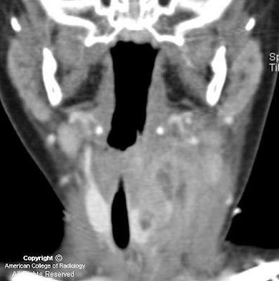
Figure 4: Neck CT coronal reformation, contrast enhanced. There is extensive phlegmon with multiloculated abscesses, extending from the left lower pharyngeal wall into the left lobe of the thyroid gland. Reactive lymphadenopathy in the left internal jugular lymph node chain is present.


Diagnosis: Infected 4th Branchial apparatus cyst


The main differential diagnostic considerations for a cystic neck mass in children include suppurative lymph nodes, abscess, thyroglossal duct cyst, lymphatic malformation, ranula, and branchial apparatus cyst. A branchial apparatus cyst (BAC) results from maldevelopment of an embryonic branchial apparatus (branchial cleft, arch, and pouch). Embryologically, 6 mesodermal branchial arches, separated by 5 external ectodermal branchial grooves (clefts) and 5 internal endodermal branchial pouches are present bilaterally. The majority of branchial apparatus anomalies are cysts that can arise from a remnant of a groove, arch, or pouch. A 2nd BAC is the most common and accounts for >90% all branchial cleft anomalies discovered in teens and adults. It represents 66%-75% of these anomalies discovered in children.

A 1st BAC is typically found as a cystic mass around the pinna or extending from external auditory canal (EAC) to the angle of the mandible. It can communicate with the external auditory canal. The 2nd BAC is typically found at or immediately caudal to the angle of the mandible, lateral to the carotid space and anteromedial to the sternocleidomastoid muscle. An associated fistulous track may extend from the cyst between the external & internal carotid arteries to the palatine tonsil. The cyst can extend to the carotid bifurcation, producing a beaked configuration, which has been called the "notch sign" and which is considered pathognomonic for a 2nd BAC. The 3rd BAC is typically found in the posterior cervical space behind the carotid sheath in the upper neck and along the anterior border of sternocleidomastoid muscle in the lower neck.

A 4th BAC is rare and seen more often in female infants. It can occur anywhere from the apex of pyriform sinus to the ipsilateral thyroid lobe. Involvement with the thyroid can be understood by noting that the thyroid gland arises from the 4th branchial arch. The most typical imaging finding of a non-infected 4th BAC is a unilocular thin-walled cyst found adjacent to or within the superior lateral aspect of the left thyroid lobe. Ninety-four percent of BACs involve the left side of the neck. These cysts normally show minimal or no peripheral contrast enhancement and no calcification. When infected, a thickened cyst wall is seen and often enhances with intravenous contrast media. Infected cysts often develop higher attenuation than noninfected cysts on CT images. Associated thyroiditis/thyroid abscess is not uncommon. An esophagram may demonstrate fistulous communication between the pyriform sinus and a 4th BAC, providing a pathway for spread of infection. Surgical resection of the cyst and its associated sinus or fistulous tract is necessary for complete cure. Medically treated or incompletely resected cysts/tracts are prone to recur.

mercoledì 13 ottobre 2010

Intraorbital Lymphatic Malformation


Findings

Figure 1: There is a multilobulated mass lesion in the retroorbital region with a fluid- fluid level.
Figure 3: There is an intraconal multilobulated mass with a fluid-fluid level and mild right globe proptosis.


Diagnosis: Intraorbital Lymphatic Malformation


Vascular lesions account for 5-20% of all orbital masses, and the two most common orbital vascular lesions are venous malformations (formerly known as cavernous hemangiomas) and lymphatic malformations (LM) ( formerly known as lymphangiomas). LMs are relatively uncommon in the pediatric population and account for only 4% of all childhood orbital masses. LMs are benign and most frequently found in the head and neck. Intraorbital LMs can arise in any orbital space, but are most commonly intraconal with frequent extraconal and preseptal expansion. Histologically, a vascular malformation can contain venous and lymphatic components, hence the name lymphaticovenous malformation. Of note, 70% of orbital lymphaticovenous malformations are associated with ipsilateral, noncontiguous, intracranial vascular abnormalities.

Intraorbital venous-lymphatic malformations are present at birth, but tend not to be discovered clinically until early childhood when they enlarge as a result of either intralesional hemorrhage or lymphoid hyperplasia and result in acute proptosis. Approximately one-half of all patients with orbital LMs also complain of limited ocular mobility. Additionally, conjunctival, facial, or oral vesicles may also be observed.

Radiologic imaging of intraorbital LMs demonstrates unencapsulated, irregular, lobulated, and multicompartmental masses. These lesions can have cystic as well as more solid components. The cystic elements of these masses commonly exhibit fluid-fluid levels as a result of intralesional hemorrhage. Additionally, LMs are frequently both pre- and postseptal and intra- and extraconal. They often display orbital expansion with irregular margins that traverse tissue planes. Ultrasound images of LMs demonstrate heterogeneous, ill-defined lesions with anechoic cystic portions and extraconal extension. On CT, these masses exhibit ill-defined borders, irregular attenuations, and variable enhancement with peripheral rim enhancement in cystic regions. Additionally, calcified phleboliths can be seen on CT in venous portions of these lesions. MR imaging is the preferred imaging modality to evaluate the location, vascular components and evolving blood products of venous-lymphatic malformations. LMs demonstrate iso- to slightly high signal intensities on T1-weighted images and very high signal intensities on T2-weighted images. MR imaging also allows for the simultaneous evaluation of the brain in an effort to detect any associated intracranial vascular anomalies.

The differential diagnosis for pediatric orbital tumors can be divided into osseous and non-osseous lesions. Pediatric osseous lesions of the orbit include dermoid inclusion cysts, which are most common, fibrous dysplasia, juvenile ossifying fibroma, osteosarcoma, Langerhans cell histocytosis, granulocytic sarcoma and neuroblastoma bone metastases. Non-osseous lesions of the pediatric orbit include most commonly rhabdomyosarcoma, but additionally infantile fibromatosis, infantile hemangioma, and LM.

Orbital lymphaticovenous malformations are histologically benign, but can demonstrate aggressive behavior, such as vision loss, as they expand. The treatment of such lesions is focused on ameliorating pain, alleviating optic nerve compression, maintaining ocular alignment and improving cosmetic appearance. Surgical resection is the preferred treatment for these lesions, and while complete removal can often be achieved with well-demarcated extraconal lesions, more diffuse intraconal lesions tend to be treated with subtotal resection.

mercoledì 22 settembre 2010

Melanotic Neuroectodermal Tumor of Infancy (MNTI)


Findings

Large mass involving left side of face with invasion, mass effect, and extensive bony involvement. The mass does not appear to cross the midline. Enhancing focus on MR adjacent to the superior aspect of the falx and superior sagittal sinus. FDG-avid spine lesions.


Diagnosis: Pathology-proven melanotic neuroectodermal tumor of infancy (MNTI)


Discussion

MNTI is a rare osteolytic, pigmented neoplasm that typically affects the head and neck, predominantly the maxilla, of infants. It is typically considered a benign lesion, with only a few reported cases of metastatic disease reported in the literature. It usually presents in the first year of life. It may appear as a rapidly expanding, non-ulcerated, lightly pigmented, blue or black lesion on the anterior aspect of the maxilla. It may extend intraorally, cause bone destruction and dislodgement of teeth.

MNTI is considered to be of neural crest origin, and some patients will high urinary excretion of vanillylmandelic acid (VMA). The tumor is typically non-encapsulated, showing local invasion of bone. The histologic appearance is similar to other cells of neural crest origin, demonstrating small, round blue cells, as well as containing a moderately vascular fibrous background. Part of the lesion may contain large polygonal cells arranged in sheets that contain melanin. Immunohistochemistry and electron microscopy can aid in the final diagnosis. The few reported cases of malignant disease have noted increased numbers of mitoses per high-powered field.

Conventional radiography may demonstrate a well-circumscribed or ill-defined radiolucency, with destruction of bone as the lesion progresses. CT can delineate the extent of soft tissue involvement and osteolysis. Contrast-enhanced MRI can demonstrate soft tissue tumors with nonenhancing, heterogeneous tissue density and can also demonstrate osseous involvement. There may be foci of T1-hyper/T2-hypointensity secondary to melanin.

Surgical excision with partial maxillectomy and 5 mm margins are typically curative, with 10-15% recurrence rates. There are no standards of care for malignant disease.

Initial biopsy of this patient's facial lesion demonstrated a high mitotic index. After 2 surgical excisions and recurrence, the patient developed intracranial and spinal column metastases. The patient underwent multiple cycles of radiotherapy and chemotherapy. The intracranial lesion was never biopsied due to the precarious location, however it has regressed, and there appears to be a slight interval decrease in size of the soft tissue component of the maxillary lesion.

giovedì 26 agosto 2010

Germinal matrix hemorrhage


Findings

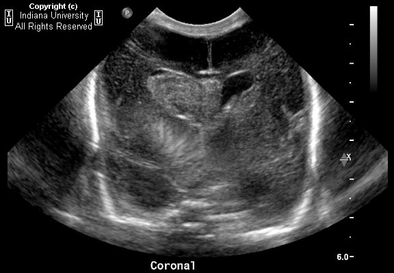
Hyperechogenic mass like hemorrhage into the right caudothalamic groove with ventricular extension, extension into the septum pellucidum, and resultant hydrocephalus; Grade 3 intraventricular germinal matrix hemorrhage.


Diagnosis: Germinal matrix hemorrhage


Key points

Risks for germinal matrix hemorrhage:
- Less than 32 weeks gestational age
- Under 1500g
- 20-25% risk in premature, low birth weight infant

Germinal matrix
- Proliferating cells; lines the periventricular spaces of the lateral ventricles and third ventricle
- Largest at 24-26 weeks, then involutes
- Begins involuting at the level of the third ventricle
- Involuted completely at 36 weeks

Mechanisms behind germinal matrix hemorrhage-
- Venous bleed
- From cardiovascular instability from lack of autoregulatory mechanism in the preterm infant
- Watershed bleed
- Nearly exclusively occurs in the first 1 wk of life, 50% on day 1
- Identifying and grading germinal matrix hemorrhage
- Blood is always as dense as choroid plexus
- Classic hemorrhage in the caudothalamic groove looks like a mass. May extend into the ventricles

Classification:
- Grade 1: Classic case caudothalamic groove looks like a mass
- Grade 2: Intraventricular extension, usually no long term sequelae
- Grade 3: Dilated ventricles
- Grade 4: Parenchyma, actually from venous hemorrhagic infarct

giovedì 19 agosto 2010

Joubert syndrome


Findings

There is agenesis of the vermis. The interpeduncular fossa is deep. There is a cerebellar cleft. There is prominence and thickening of the superior cerebellar peduncle. There is a bat wing configuration of the fourth ventricle.


Diagnosis: Joubert syndrome


Key points

Joubert syndrome is an autosomal recessive genetic disorder that affects the cerebellum and midbrain. There is agenesis/hypogenesis of the cerebellar vermis and malformation of the midbrain and brainstem.

With poor development of the cerebellar vermis and brainstem, patients can present with ataxia, hypotonia, and hyperpnea. One of the first clinical signs of Joubert is episodic hyperpnea or apnea in an infant. Breathing can reach upwards of 160 breaths/minute. Mild mental retardation is often seen, though some may have normal intelligence. Patient health and growth are usually not affected. Patients with Joubert benefit from physical, occupational, and speech therapy. Infants with respiratory abnormalities require respiratory monitoring.


Radiologic overview of the diagnosis

The initial radiologic manifestations are often seen on fetal ultrasound with increased nuchal lucency being the most apparent finding. Fetal or Brain MR is the best modality to evaluate the presence of Joubert syndrome.

The classic findings are a hypoplastic/aplastic vermis, thick superior cerebellar peduncles, a deep interpeduncular fossa, and an abnormal midbrain. This configuration resembles a molar tooth and is often referred to as the "molar tooth" sign. The fourth ventricle also takes on a bat wing configuration. The midbrain is usually decreased in AP dimension. In this case, there is complete agenesis of the vermis and the molar tooth sign can be seen with thickening of the superior cerebellar peduncles. The corpus callosum and pars intermedia is thickened and may reflect a compensatory response by the brain adapting to the lack of a vermis.