martedì 28 febbraio 2006

Moya moya disease


Findings

There is an area of increased attenuation with surrounding edema in the posterior limb of the left internal capsule, extending into the corona radiata. This represents acute hemorrhage.
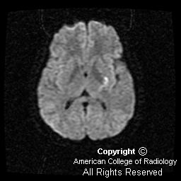
Diffusion-weighted imaging demonstrates the magnetic susceptibility of the acute hemorrhage (Figure 4). However, inferior to the hemorrhage are areas of increased signal distinct from the hemorrhage, suggesting associated infarction (Figure 2 and Figure 3).
MIP image from the Circle of Willis study demonstrates bilateral supraclinoid carotid artery occlusion (Figure 5) There is no flow in the anterior cerebral artery territory, minimal flow in the middle cerebral artery territory, and hypertrophy of the posterior circulation (Figure 5).
IVI image of the 3D TOF Circle of Willis study again demonstrates the presence of bilateral supraclinoid carotid artery occlusion (Figure 6).
A single source image from the Circle of Willis MRA demonstrates the classic hypertrophy of the lenticulostriate arteries seen in Moya moya (Figure 7).
This single lateral view from catheter angiogram of the right internal carotid artery, performed for evaluation of possible treatment options, better demonstrates the classic finding of the "puff of smoke," caused by the hypertrophied lenticulostriate vessels (Figure 8).


Diagnosis: Moya moya disease


Translated literally as “hazy puff of smoke” in Japanese, Moya moya is a chronic cerebrovascular occlusive disease involving the Circle of Willis. The pathologic findings of Moya moya include intimal thickening and internal elastic lamina disruption of the arteries of the Circle of Willis and collateral vessels. The angiographic features of the disease include bilateral stenosis or occlusion of the supraclinoid portion of the internal carotid artery that extends to the proximal portions of the anterior cerebral artery and middle cerebral artery, as well as the presence of parenchymal, leptomeningeal, or transdural collateral vessels that supply the ischemic brain. These collateral vessels are typified by a meshwork appearance. Aneurysm formation is quite common, most often affecting the anterior communicating and anterior cerebral arteries with unilateral Moya moya, and basilar artery with bilateral Moya moya. Extracranial involvement of Moya moya has been described in the literature, most frequently involving the renal arteries.

Cerebral angiography is still the gold standard for diagnosis of Moya moya, and it is using this modality that we can best visualize the appearance of these collateral vessels as a “puff of smoke.” CT is useful for diagnosing acute hemorrhage or prior infarct. MR imaging has also been widely used in assessing anatomic detail and the vascular architecture. MR often demonstrates loss of distal internal carotid artery flow void, as well as small punctate hypointense lesions on T1-weighted imaging in the basal ganglia. These punctate hypointense lesions are the collateral vessels that form due to the occlusion of larger vessels, such as the Circle of Willis.

Moya moya commonly afflicts people of Japanese and Asian decent, but has been diagnosed in people of different races and ethnicities. It has a slight predilection for females (1.8:1) and affects both children and adults. There is increased mortality in adults compared to children (10%:4.3 % respectively); and in greater than 50% of all patients, the disease results in progressive deterioration of cognitive function. The etiology of Moya moya is unknown, but associations with conditions such as sickle cell disease, neurofibromatosis, Down’s syndrome, and tuberculosis have been documented in the literature.

The clinical features of Moya moya can be divided by age group (adult and pediatric) or presenting symptom (epilepsy, ischemic events, or hemorrhagic stoke). Epilepsy and ischemic events are more common in children, whereas intracerebral hemorrhage is far more common in adults. Hemorrhage is most common in the thalamus, basal ganglia, and frontal lobe tissue adjacent to the frontal horn of the lateral ventricles. Treatment of acute Moya moya is usually symptomatic, and often involves surgery. Options include direct and indirect revascularization. Medical treatment is given as the presenting symptoms dictate.

Differential diagnosis of Moya moya includes carotid thromboembolic disease, blood dyscrasias and stroke, and vasculitidies such as polyarteritis nodosa.

mercoledì 15 febbraio 2006

Allergic fungal sinusitis


Findings

Axial CT image reveals erosive changes (Figure 1).
Axial CT image (Figure 2) demonstrates characteristic expansion of the ethmoid air cells (Figure 2). Hyperdense material is also located in the sphenoid sinus centrally within the sinus and is surrounded by hypodense mucosa (Figure 2).
Coronal CT image again reveals similar findings including erosion and expansion (Figure 3)


Diagnosis: Allergic fungal sinusitis


Allergic fungal sinusitis (AFS) is believed to be an allergic reaction to aerosolized environmental fungi in an immunocompetent host. This process is quite different than that of the invasive fungal infections seen in immunocompromised patients, such as those with diabetes mellitus or AIDS. The pathologic process may be a Type 1, IgE-mediated hypersensitivity reaction to the presence of fungus. Additional factors such as anatomy, genetics, and possible lymphocyte sensitization may also be involved. Most patients have a history of allergic rhinitis, but timing of the development of the process is varied. Thick fungal debris and mucin develop within the sinuses and must be excised in order to remove the inciting allergen.

AFS usually involves multiple sinuses and can be unilateral or bilateral. The ethmoid sinus is the most common location, followed by the maxillary, frontal, and sphenoid sinuses. Expansion and even erosion of the sinuses is characteristic. In fact, there is approximately a 20% rate of erosion with extension into adjacent cavities. Sites of extension include the nasopharynx and pterygomaxillary space, as well as the intracranial and intraorbital regions. The sinuses are usually opacified by centrally hyperdense material and a peripheral rim of hypodense mucosa. Hypointensity on T1-weighted imaging and T2-weighted imaging is the most common finding on MRI. The peripheral mucosa demonstrates enhancement on contrasted images.

AFS is usually treated by local excision and steroid therapy. Antifungal therapy is also attempted in some cases, but results of this therapy are equivocal. Recurrence after surgery is not uncommon; however, the inclusion of steroid therapy significantly reduces recidivism.

martedì 14 febbraio 2006

Posterior pituitary ectopia


Findings

The posterior pituitary bright spot (Figure 2 and Figure 3) is ectopic, located just inferior and posterior to the tuber cinereum. The adenohypophysis is small. The pituitary stalk (Figure 2 and Figure 3) is hypoplastic and difficult to see.


Diagnosis: Posterior pituitary ectopia


Posterior pituitary ectopia is a developmental disorder of the porencephalon, thought to be genetic in origin. Multiple genes have been implicated. The end result is defective neuronal migration. Patients often have other midline abnormalities, such as septooptic dysplasia and lobar holoprosencephaly. Other structures developing at the same time may be affected. The optic/olfactory nerves and the anterior pituitary should be carefully evaluated, as they may be abnormal or absent. Additional associated syndromes include: Kallmann sydrome, CHARGE syndrome, and Pallister-Hall syndrome. Patients are imaged because of other abnormalities, hypopituitarism, and short stature. The anterior pituitary may be affected by absence or hypoplasia of the infundibulum.

Cranial computed tomography may reveal the pituitary fossa to be small. On MRI, the posterior pituitary bright spot (bright secondary to the accumulation of ADH-neurophysin) is ectopic and often seen just below the tuber cinereum. The ectopic pituitary is hyperintense on T1-weighted imaging. The adenohypophysis is small. On either CTA, conventional angiography, or MRA the supraclinoid carotid arteries approach the midline and appear to be “kissing.” The frontal lobes may be abnormal.

venerdì 10 febbraio 2006

Spinal involvment in Hodgkin's lymphoma


Findings

Sagittal T1 image (Figure 1) in a patient with Hodgkin's lymphoma. Compression deformity of the C4 vertebral body is noted and there is abnormal marrow signal. There is an associated mass with epidural extension of tumor that directly impinges on the cord, displacing intervening CSF.

Two sagittal T2 images (Figure 2 and Figure 3) in a patient with Hodgkin's lymphoma show compression deformity of the C4 vertebral body (Figure 2). There is an associated mass with epidural extension of tumor that directly impinges on the cord with additional tumor seen anterior to the vertebral body. Displacement of the intervening CSF is better demonstrated on this T2-weighted sequence (Figure 3).

Axial T2 image (Figure 4) at the affected level confirms that the anterior aspect of the cervical canal is flattened by the tumor with loss of intervening CSF and deformity of the cord. These findings are indicative of cord compression.


Diagnosis: Spinal involvment in Hodgkin's lymphoma


Acute spinal cord compression is a potentially devastating neurological emergency that requires both prompt diagnosis and intervention to prevent permanent impairment. The frequency of metastatic cord compression is increasing as cancer prevalence rises and new treatment modalities prolong patient survival. Approximately 5% of patients with terminal cancer develop epidural spinal cord compression. Metastases are 25 times more common than primary tumors as a causative etiology. Intramedullary spinal cord metastasis have frequency of 1/16 that for epidural metastasis and are best diagnosed by MRI. Of note, approximately 20% of cancer patients with spinal cord compression have the associated new neurological deficits as their initial manifestation of disease.

Many types of tumor metastasize to the epidural space. Relative incidence of spinal cord compression by a particular type of tumor is determined by a combination of tumor prevalence in the population and its predilection for spinal involvement. The most common tumors causing cord compression in adults are prostate, breast, lung, NHL, multiple myeloma, renal, and colorectal cancer. The most common types in children are germ cell tumors, Hodgkin's, and sarcomas, especially Ewing’s.

Magnetic resonance imaging is the study of choice in the evaluation of suspected cord compression as it is noninvasive, does not involve radiation, and provides for investigation of both osseous and soft tissue lesions. Whole spine imaging is generally undesirable as it is more time-consuming, expensive, and difficult for patients who are often in considerable pain. It further lowers resolution on exams that are often suboptimal secondary to severe patient pain and patient movement.

Therefore, information from the neurological exam is critical for localization of the lesion and optimization of the imaging protocol. Spinal sensory levels on neurological examination may be up to several segments below the anatomic level of cord compression. Evaluation of motor function and reflexes is very useful for lesion localization. Pain is ubiquitous in cancer patients, and while it may be initially localized, it is not specific to cord compression and is more often related to vertebral metastasis and pathologic fracture. Pain increases in intensity with worsening compression. Pain from cord compression often worsens with recumbency.

Once the site of interest is more precisely identified, sagittal T1 and T2 images and axial T2 images are required for the diagnosis. Spinal cord compression may be defined on imaging as the presence of a mass lesion abutting the cord with the complete loss of intervening CSF. This must be accompanied by deformation of the spinal cord or the presence of signal changes within the cord. The findings are best visualized on T2-weighted images. If the patient is concurrently symptomatic, acute intervention is mandated with the specific type of intervention determined by the underlying disease process.

martedì 7 febbraio 2006

Pars interarticularis stress fracture


Findings

CT images demonstrate lucencies through the pars bilaterally (yellow arrows in Figure 4) involving the anterior and posterior cortical margins on the left (Figure 6) and anterior cortical margin on the right (Figure 5). The irregular margins suggest recent injury. There was no evidence of listhesis on sagittal reconstructions.
T2 fat saturated MR images demonstrate edema in the pars and pedicles bilaterally (Figure 7, Figure 8, and Figure 9).


Diagnosis: Pars interarticularis stress fracture


Asymptomatic spondylolysis and spondylolisthesis occur in 6%-8% of the general population and are often identified incidentally. A percentage of individuals who are genetically predisposed to developing these entities will become symptomatic with or without an inciting injury. A high prevalence among athletes who participate in activities that subject the lumbar spine to repetitive loading suggests that trauma is also an etiologic factor. Spondylolysis and spondylolisthesis involving the lumbar spine are the most frequently diagnosed organic causes of back pain in children and adolescents). The child or adolescent typically presents with low back pain occasionally radiating to the buttocks or posterior thigh. The pain is usually insidious in onset and progressively increases in severity. The diagnostic work varies widely by clinician.

PA and lateral radiographs of the lumbosacral spine are used as a screening evaluation and can assess overall spinal alignment. They may also identify focal sclerosis associated with stress reactions of the pars, lamina or pedicle, as well as defects of the pars. Single-photon emission computed tomography (SPECT) of the spine is indicated in the evaluation of patients with inconclusive radiographs but whose history and clinical examination suggest the presence of spondylolysis. Increased uptake in the pars, adjacent lamina, or pedicle - either unilateral or bilateral - suggests a stress reaction, stress fracture, or symptomatic spondylolytic defect. SPECT, however, may be positive in other etiologies, including neoplasm and acute fracture. A negative SPECT in the presence of a spondyloltic defect identified on radiographs is an incidental finding.

CT is often used to define the bone morphology of spondylolysis. Stress reaction is defined as localized sclerosis without trabecular or cortical disruption of the pars, lamina, or pedicle. Stress fracture is defined as cortical or trabecular disruption of the pars with minimal sclerosis or lysis of the fracture gap. Pars disruption with surrounding sclerosis and a definable area of lysis at the site of the pars discontinuity is diagnostic of a spondylolytic defect.

Although the overall role of MRI has yet to be determined, MRI may also be obtained to identify marrow edema at the pars or if neurologic symptoms are present. Stress injuries to the lumbar pars interarticularis initially manifest as bone marrow edema visible as abnormal high T2 signal on sagittal fat-saturated MR images. Signal abnormalities may also be seen in the adjacent pedicle and articular process. As the stress injury progresses, thinning, fragmentation, or irregularity of the pars interarticularis may become visible.

lunedì 6 febbraio 2006

Juvenile pilocytic astrocytoma


Findings

Figure 1: Noncontrast CT demonstrates a large midline posterior fossa solid and cystic mass with associated compression of the fourth ventricle.
Figure 2: Axial FLAIR demonstrates a solid nodule and a tumor cyst. Surrounding vasogenic edema is present.
Figure 3: Axial post-contrast T1 MR image demonstrates diffuse enhancement of the solid tumor nodule.
Figure 4: Sagittal post-contrast T1 image demonstrates cerebellar tonsillar herniation.


Diagnosis: Juvenile pilocytic astrocytoma


Primary brain tumors are the most common solid neoplasms in children, representing approximately 20% of all pediatric tumors. The posterior fossa is the most common location of primary brain tumors in children.

The most frequent posterior fossa tumors in children are juvenile pilocytic astrocytoma (JPA), medulloblastoma, ependymoma, and brainstem glioma. The JPA is second in frequency only to medulloblastoma.

Pilocytic astrocytomas usually present within the first two decades of life, with a peak age of presentation at 10 years. Patients present with headache, nausea, and vomiting, usually secondary to hydrocephalus, or may have symptoms because of brainstem or cerebellar compression.

When completely removed, there is a greater than 90% five-year survival rate. Surgery is usually curative, but prognosis does depend on cellular morphology.

The typical radiographic appearance is that of an enhancing mural nodule, as well as a nonenhancing cystic component. Most are well-defined and located off the midline in the cerebellar hemispheres or vermis. The nodular portion of the lesion usually demonstrates homogenous contrast enhancement. Calcification is uncommon. Twenty per cent are solid without cysts. MRS can demonstrate elevated choline, lactate, and myoinositol. Avid uptake is often present on PET scans.