Visualizzazione post con etichetta Cysts. Mostra tutti i post

lunedì 22 novembre 2010

Suprasellar arachnoid cyst


Findings:

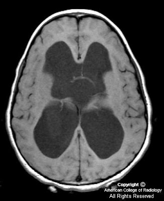
Figure 1, Figure 2 and Figure 3 show severe hydrocephalus involving the lateral and third ventricles.
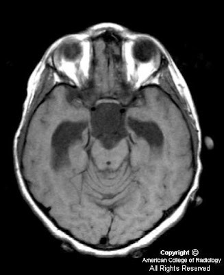
Figure 4, Figure 5, Figure 6, show a suprasellar mass lesion which follows CSF signal on all sequences, including FLAIR.


Diagnosis: Suprasellar arachnoid cyst


Arachnoid cyst is the most common congenital lesion of the brain. It is typically an incidental imaging finding and is rarely symptomatic. However, in this case the size and location of the lesion in the suprasellar cistern resulted in severe obstructive hydrocephalus and precocious puberty. Both of these complications resolved following surgical drainage of the cyst.

Arachnoid cysts arise from a splitting of the arachnoid membrane with formation of a cyst wall consisting of fibrous connective tissue. There is no epithelial lining in the wall. Expansion occurs following trapping of cerebral spinal fluid through defects in the cyst wall. Arachnoid cysts occur most commonly in the middle cranial fossa and have a 4-to-1 male-to-female ratio. Even large cysts tend to be asymptomatic. Associated clinical features in symptomatic patients include headache, calvarial bulging, intracranial hypertension, craniomegaly, developmental delay, visual loss, precocious puberty, and seizures. Treatment of arachnoid cysts is not recommended by many unless there is a clear cause and effect relationship between the cyst and symptoms as shown in this case.

MRI is the preferred diagnostic modality for arachnoid cysts because of its ability to demonstrate the location, extent and relationship of the cyst to surrounding neurologic structures. Lesions typically have the signal intensity of CSF on all sequences, do not enhance and do not demonstrate restricted diffusion. The most important differential diagnostic consideration is between arachnoid and epidermoid cysts. Epidermoid cysts show restricted diffusion on diffusion-weighted images. In addition, unlike epidermoid cysts, arachnoid cysts show suppressed signal on fluid-attenuated inversion recovery (FLAIR) images.

lunedì 25 ottobre 2010

Infected 4th Branchial apparatus cyst


Findings

Figure 1: Neck CT, contrast enhanced, at level of pyriform sinus. The left pyriform sinus is effaced by an inflammatory mass.
Figure 2: Neck CT, contrast enhanced, at level of subglottic trachea. The image demonstrates continuation of the large inflammatory mass with small areas of necrosis or abscesses. Note displacement of the trachea to the right and lateral displacement of the carotid sheath vessels. Reactive lymphadenopathy is present in the internal jugular chain.
Figure 3: Neck CT, contrast enhanced, at level of the thyroid gland. The image shows a mixed attenuation mass in the enlarged left lobe of the thyroid. This mass arises from extention of the extrinsic anterior and lateral inflammatory mass with phlegmon and abscesses from an infected 4th branchial apparatus cyst.
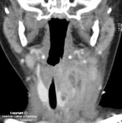
Figure 4: Neck CT coronal reformation, contrast enhanced. There is extensive phlegmon with multiloculated abscesses, extending from the left lower pharyngeal wall into the left lobe of the thyroid gland. Reactive lymphadenopathy in the left internal jugular lymph node chain is present.


Diagnosis: Infected 4th Branchial apparatus cyst


The main differential diagnostic considerations for a cystic neck mass in children include suppurative lymph nodes, abscess, thyroglossal duct cyst, lymphatic malformation, ranula, and branchial apparatus cyst. A branchial apparatus cyst (BAC) results from maldevelopment of an embryonic branchial apparatus (branchial cleft, arch, and pouch). Embryologically, 6 mesodermal branchial arches, separated by 5 external ectodermal branchial grooves (clefts) and 5 internal endodermal branchial pouches are present bilaterally. The majority of branchial apparatus anomalies are cysts that can arise from a remnant of a groove, arch, or pouch. A 2nd BAC is the most common and accounts for >90% all branchial cleft anomalies discovered in teens and adults. It represents 66%-75% of these anomalies discovered in children.

A 1st BAC is typically found as a cystic mass around the pinna or extending from external auditory canal (EAC) to the angle of the mandible. It can communicate with the external auditory canal. The 2nd BAC is typically found at or immediately caudal to the angle of the mandible, lateral to the carotid space and anteromedial to the sternocleidomastoid muscle. An associated fistulous track may extend from the cyst between the external & internal carotid arteries to the palatine tonsil. The cyst can extend to the carotid bifurcation, producing a beaked configuration, which has been called the "notch sign" and which is considered pathognomonic for a 2nd BAC. The 3rd BAC is typically found in the posterior cervical space behind the carotid sheath in the upper neck and along the anterior border of sternocleidomastoid muscle in the lower neck.

A 4th BAC is rare and seen more often in female infants. It can occur anywhere from the apex of pyriform sinus to the ipsilateral thyroid lobe. Involvement with the thyroid can be understood by noting that the thyroid gland arises from the 4th branchial arch. The most typical imaging finding of a non-infected 4th BAC is a unilocular thin-walled cyst found adjacent to or within the superior lateral aspect of the left thyroid lobe. Ninety-four percent of BACs involve the left side of the neck. These cysts normally show minimal or no peripheral contrast enhancement and no calcification. When infected, a thickened cyst wall is seen and often enhances with intravenous contrast media. Infected cysts often develop higher attenuation than noninfected cysts on CT images. Associated thyroiditis/thyroid abscess is not uncommon. An esophagram may demonstrate fistulous communication between the pyriform sinus and a 4th BAC, providing a pathway for spread of infection. Surgical resection of the cyst and its associated sinus or fistulous tract is necessary for complete cure. Medically treated or incompletely resected cysts/tracts are prone to recur.

venerdì 1 ottobre 2010

Tornwaldt cyst


Findings

There is no intracranial hemorrhage, calvarial fracture or transtentorial herniation. On the lower images of the brain, there is a cystic hypoattenuating lesion at the posterior wall of the nasopharynx measuring 1.4 x 1.2 cm.


Diagnosis: Tornwaldt cyst


A Tornwaldt cyst is a benign proteinaceous cyst that is located in the midline of the posterior nasopharynx, superficial to the superior constrictor muscle of the pharynx. It is surrounded by adenoid tissue and arises from notochordal remnants in the pharyngeal bursa (pouch of Luschka). They are seen in up to 4% of the population (equally in males and females) and are usually asymptomatic unless they become infected. If infected, they can cause a variety of symptoms including purulent drainage, sore throat, prevertebral muscle spasms, halitosis and Eustachian tube obstruction.

These fluid-filled cysts are usually discovered on imaging (both CT and MRI) as incidental findings. The cyst is well-circumscribed and located in the midline in the posterior nasopharynx (between the longus coli muscles). On CT, it is hypo-attenuating and appears cystic. It will almost invariably appear bright on T2-weighted images. The T1 signal will vary from CSF signal to very bright hyperintensity depending on the amount of protein, fat, hemorrhage and mucus within the cyst. A thin rim of peripheral enhancement may be seen with gadolinium administration. Nasopharyngoscopy, although not necessary for asymptomatic cases, can help supplement the diagnosis.

Treatment is not necessary in most cases. For the rare symptomatic cases treatment options include surgical excision, electrocoagulation or marsupialization.

giovedì 23 settembre 2010

Filar cyst


Findings

Figure 1: Axial T2-weighted image demonstrates a cystic structure in the filum terminale.
Figure 2 and Figure 3: Coronal T2-weighted images demonstrate a cystic structure in the proximal filum terminale.


Diagnosis: Filar cyst


The human spinal cord develops through three distinct stages: neurulation, canalization, and retrogressive differentiation. The conus medullaris, filum terminale, and cauda equine are mainly developed and formed in the retrogressive differentiation stage as the caudal cell mass regresses.

Cystic structures within the distal spinal cord, conus medullaris, and filum terminale are commonly seen on routine lumbosacral spine sonography in the neonates. These patients often present with abnormal laboratory values and/or external body features that are suggestive of underlying spinal dysraphism or neural tube defects. The diagnostic consideration of a cystic lesion in this region includes: syrinx, neoplasm (ependymoma or astrocytoma), persistent ventriculus terminalis in the conus medullaris, and filar cyst in the filum terminale of the cord.

Filar cysts are a relatively common entity in the neonates usually detected on screening lumbosacral sonograms. However, it has not been extensively reported in the literature. It is considered a normal variant when found as an isolated finding. The exact etiology of a filar cyst has not been reported. Literature has suggested that filar cysts are developmentally similar to the septum pellucidum and ventriculus terminalis, which can regress with age. On ultrasound, it is usually describes as an anechoic, cystic structure completely contained within the filum terminale. MRI of the lumbosacral spine can be obtained in cases that are questionable on ultrasound. Filar cyst follows the typical characteristic of a simple cyst in all sequences of an MR study. Ventriculus terminalis is a normal developmental variant described as a nonenhancing dilation of the ependyma-lined central canal at the level of the conus medullaris. Persistent ventriculus terminalis deserves special attention such that it is often used interchangeably with filar cyst in the literature, in the setting of cystic lesions seen in the distal lumbar spinal cord proximal to the conus medullaris. A cystic lesion in the absence of a solid component makes a neoplastic process less likely. A syrinx isolated to the distal spinal cord is also less common as it usually has a superior extension.

Treatment options are primarily based on patient's symptomatology. In asymptomatic neonates/infants, no further imaging is needed.

giovedì 14 gennaio 2010

Arachnoid cyst with complicating intracystic and subdural hemorrhage


Findings

Figure 1 and Figure 2: Initial non-contrast (Figure 1) and contrast enhanced (Figure 2) axial head CT images. At the level of lateral ventricles an isoattenuating left sided subdural hematoma with mass effect and midline shift is demonstrated. Contrast enhanced imaging shows no abnormal enhancement within the subdural hematoma.
Figure 3 and Figure 4: More inferiorly, a hemorrhagic round extra-axial mass in anterior aspect of the left middle cranial fossa is seen. Bone window image (Figure 4) demonstrates focal thinning with endosteal scalloping of the overlying bone.

Figure 5: Diffusion weighted image through the lesion in the left middle cranial fossa shows no evidence of abnormally restricted diffusion of water. This confirms the lesion is not an epidermoid cyst as they characteristically show intense signal abnormality on DWI.
Figure 6, Figure 7, Figure 8, and Figure 9: The lesion demonstrated isointense signal on T2-weighted imaging (Figure 6). T1-weighted MR imaging (Figure 7) better demonstrates the lesion's complex nature. The lesion represents an arachnoid cyst which shows evidence of hemorrhage as demonstrated by the presence of T1 shortening. The surrounding subdural hematoma shows well demarcated prominent T1 shortening indicative of hemorrhage. Contrast enhanced axial T1-weighted MR imaging (Figure 8) shows no enhancement within the arachnoid cyst but does show enhancement of the dural membrane (arrow). The coronal contrast enhanced T1-weighted MR image (Figure 9) shows the arachnoid cyst, arachnoid membrane, and subdural hematoma.


Diagnosis: Arachnoid cyst with complicating intracystic and subdural hemorrhage


The widespread use of neuroimaging has increased incidental detection of arachnoid cysts. Arachnoid cysts constitute approximately 1% of all intracranial space-occupying lesions. Common locations include the middle cranial fossa, cerebral convexity, perisellar, retrocerebellar, cerebellopontine angle, and quadrigeminal plate cisterns. While some arachnoid cysts arise from post-inflammatory changes after trauma, intracranial hemorrhage, or infection, most are thought to be congenital. It is theorized that these cysts form due to an aberration in arachnoid development resulting in splitting or duplication of the membrane, a defect in condensation of the mesenchyme, or abnormalities of CSF flow. The exact means of arachnoid cyst genesis remains unclear.

On both CT and MRI scans arachnoid cysts are non-enhancing well-circumscribed extra-axial lesions with the same attenuation (on CT) or signal (on all MRI pulse sequences) as that of CSF. Bony erosion and remodeling, features suggestive of longstanding processes, are often associated with arachnoid cysts. These findings are seen in nearly half of cases, and are felt to be secondary to chronic fluid accumulation with transmitted CSF pulsations. Adjacent deformity or even hypoplasia of the subjacent brain may be present depending on the size and location of the cyst.

Rare complications of arachnoid cysts such as intracystic hemorrhage, subdural hematoma, or subdural hygroma may occur either after head injury or spontaneously. Rupture of the outer arachnoid cyst wall and surrounding fragile veins allows blood to accumulate within the cyst and/or subdural compartment. Intracystic hemorrhage can also be due to interruption of the highly vascular arachnoid cyst membrane or of the bridging veins traversing the cyst cavity. Fluid production by flat arachnoid cells lining the cyst walls may explain spontaneous cyst enlargement and hemorrhage if sufficient intracystic pressure is attained to breech the wall and disrupt the vasculature.

lunedì 28 dicembre 2009

Colloid cyst


Findings

The CT of the paranasal sinus shows a circumscribed hyper dense homogenous mass at midline anterior to third ventricle. The MRI of the brain shows an approximately 1 cm circumscribed mass location as above. Isointense on T1. Iso/slight hyper intense on T2. Hyperintense on FLAIR. No diffusion restriction or enhancement. Some heterogeneity of inferior margin (seen on sagittal). There is hydrocephalus of the left lateral ventricle.

Differential diagnosis:
- Colloid cyst
- Pseudocyst (CSF flow artifact)
- Choroid plexus papilloma
- Glioma
- Hamartoma
- Neurocysticercosis
- Granulomatous disease


Diagnosis: Colloid cyst


Colloid cyst

Not a true neoplasm; rather a remnant from embryonic endoderm
Contains variable contents including mucinous secretions and desquamated epithelial cells. This variability accounts for variable MRI signal characteristic
Usually present in 3rd to 4th decade
2% of all intracranial masses, 15-20% of all intraventricular masses
Only 10% show growth
Classic clinical scenario is acute onset headache (duet to acute obstruction and hydrocephalus) which is reproduced with tilting head forward (Brun phenomenon)
Fatalities reported due to this lesion (though not common)


Radiologic overview of the diagnosis

The location of this lesion is nearly pathognomonic = Attached to anterior/superior 3rd ventricular roof, wedged into foramen of Monro, with pillars of fornix draped around cyst.
Variable size at presentation, mean size = 15 cm
May be associated with ventriculomegaly

CT:
- 2/3 hyper dense, 1/3 iso- to hypo- dense (dependent on hydration state)
- Usually no enhancement, rarely rim enhances

MRI:
- T1WI usually hyper intense (2/3), 1/3 isointense
- T2WI majority isointense, but more variable, reflects hydration content of internal materials
- Can see fluid-fluid levels
- FLAIR: Does not suppress (hyper intense)
- DWI: No restriction
- Rare rim enhancement

lunedì 14 settembre 2009

Spinal Arachnoid Cyst


Findings

Figure 1: Sagittal T1 FLAIR image shows subtle distortion of the anatomy at the conus and proximal cauda equina. No mass is seen.
Figure 2: Sagittal T2 weighted image shows subtle distortion of the anatomy at the cauda equina. There is a suggestion of a T2 hyperintense lesion splaying the roots of the cauda equina.
Figure 3: Axial T2 weighted image shows centrifugal displacement of the nerve roots of the cauda equina. There is T2 hyperintense material, similar in signal to CSF, centrally.
Figure 4: PA and lateral views from a contrast myelogram following introduction of 10 cc of Isovue 200M into the subarachnoid space shows six lumbar vertebral bodies and a complete myelographic block by a convex bordered mass at L2-3.
Figure 5: 20-minute delayed sagittal and coronal reformatted images from CT-myelogram shows the almost round cyst at L2 causing an incomplete block (there is a mild amount of contrast above the cyst). Note the cyst contents do not communicate with the sub-arachnoid space and hence the cyst remains low density (dark). The cyst is splaying the nerve roots of the cauda equina and posteriorly displacing the filum terminale tethering the conus medullarus posteriorly.


Diagnosis: Spinal Arachnoid Cyst


Spinal arachnoid cysts are uncommon and may be extramedullary-intradural or epidural. They are CSF fluid sacs contained by arachnoid. Clinical presentation is variable including pain, weakness, numbness, claudication, myelopathy or bladder/bowel incontinence. Symptoms may be exacerbated with postural changes and Valsalva maneuver. Many are asymptomatic incidental findings on imaging studies done for other reasons. The etiology of these cysts is debated.

Epidural cysts are most commonly located posteriorly and displace the dura. Larger lesions may cause symptoms by compressing the spinal cord. Most occur in the thoracic spine posteriorly. There may be erosion of the adjacent bony elements of the spinal canal. Synovial cysts are not arachnoid cysts but appear as epidural cysts that can be characterized because of their proximity to diseased facet joints and thick walls.

Extramedullary intradural cysts are even less common than epidural cysts and are mostly located posteriorly in the spine. They can be classified as primary or associated with trauma, infection or subarachnoid hemorrhage. Extramedually intradural cysts are difficult to visualize on MRI and CT because of their similarity in signal intensity or density of the cyst fluid to CSF. Since these cysts to do not enhance, intravenous contrast is not helpful. Discovery of these lesions primarily relies on identification of displacement of adjacent structures. Extramedullary cysts located anteriorly are even less common. Cine-MRI has been reported to be helpful for diagnosis. In this case however, CSF flow study did not reveal differential flow between the cyst and the subarachnoid spaces above or below the cyst. The cyst could not be distinguished from CSF on a multi-echo sequence (TR 2000 TE 16,32,48,64, not shown.) Most subarachnoid cysts communicate with the subarachnoid space. In this case, communication was not demonstrated on the delayed CT-myelogram (shown above) which did not reveal contrast penetration into the cyst. Rarely, a hydatid cyst could mimic an arachnoid cyst, and may be suspected when the cyst wall is markedly hypointense on T1 and T2 weighted images.

Intramedullary cysts can be easily differentiated from intradural cysts. CT may demonstrate a low density cystic cavity, with or without cord enlargement. Intramedullary cysts are easily visualized on MRI because of the differential signal intensity between cyst and surrounding spinal cord. Intravenous gadolidium based contrast agent may be helpful in cases related to neoplasia. Intramedullary cysts may be congenital, benign, post traumatic, or neoplastic. Spinal intradural cysts related to trauma or surgery can be associated with an intramedullary cyst.

Conservative management is recommended for cysts found incidentally. Symptomatic intradural cysts are treated with surgical excision. An important component of surgical treatment of epidural cysts includes closure of the arachnoid defect that is the source of the CSF leak.

lunedì 13 aprile 2009

Interhemispheric cyst


Findings

There is agenesis of the corpus callosum. There is a multiseptated, left parasagittal midline cyst. Possible communication with the third ventricle. No associated enhancement is present. The left lateral ventricle appears dilated. No midline shift is present.

Differential diagnosis:
- Interhemispheric cyst
- Holoprosencephaly


Diagnosis: Interhemispheric cyst
At surgery: No definite communication with the ventricular system was found at surgery, thus categorizing the lesion as an interhemispheric cyst – type 2.


Key points


The association between an interhemispheric cyst and agenesis of the corpus callosum has been reported, although the etiology of the cyst is uncertain (arachnoid cyst, neuroepithelial cyst, or extensions of the ventricular lining).
Classification was most recently described by Barkovich et al based on appearance and communication with the ventricular system in 25 patients. Type 1 cysts communicate with the ventricular system while Type 2 cysts do not. Lack of communication with the ventricles was determined by visualizing a wall of the loculated cyst on MRI.

Classification of agenesis of the corpus callosum with interhemispheric cyst:
- Type 1a:
Cyst: Isointense to CSF (MR), unilocular.
Communication with lateral ventricles only.
Macrocephaly, hydrocephalus, Dandy-Walker malformation.
Males.

- Type 1b:
Cyst: Isointense to CSF (MR), unilocular.
Communication with and obstruction of third ventricle.
Macrocephaly, thalamic fusion without subcortical heterotopia.
Males > Females.

- Type 1c:
Cyst: Isointense to CSF (MR), unilocular.
Communication with lateral and third ventricles.
Microcephaly, cerebral dysplasia or hypoplasia.
Males.

- Type 2a:
Cyst: Isointense to CSF (MR), multilocular.
No communication with lateral or third ventricles.
Macrocephaly, hydrocephalus.
Male.

- Type 2b:
Cyst: Hyper attenuation (CT), hyper intense (T1W MR), multilocular.
No communication with lateral or third ventricles.
Aicardi syndrome, subependymal heterotopia, polymicrogyria, seizures, hypoplastic falx cerebri, uni- or bilateral ventriculomegaly, developmental delay.
Female.

- Type 2c:
Cyst: Isointense to CSF (MR), multilocular.
No communication with lateral or third ventricles.
Subcortical heterotopia, developmental delay.
Male.

martedì 30 ottobre 2007

Arachnoid cyst with acute subdural hemorrhage


Findings

Large left frontal cystic lesion with thin septations and no enhancement. Unclear whether this lesion is intra-axial or extra-axial. No diffusion restriction (image not shown), no surrounding vasogenic edema. Also present is an acute right subdural hematoma caused by the patient's fall.

Differential diagnosis for the cystic lesion:
- Arachnoid cyst
- Ependymal cyst
- Glial cyst


Diagnosis: Arachnoid cyst. Acute subdural hemorrhage.


Key points

Arachnoid cysts are CSF containing cysts which are intra-arachnoid, and do not communicate with the ventricular system.
50-60% occur within the middle cranial fossa.
May arise as a developmental anomaly, or may be acquired as a complication of adhesions.
Small number are associated with neoplasms.
Walls of cyst are formed by splitting of the arachnoid membrane.
Usually asymptomatic. Can have headache or seizures.
The most effective surgical treatment appears to be excision of the outer cyst membrane and cysto peritoneal shunting.

venerdì 27 luglio 2007

Arachnoid cyst with hemorrhage


Findings

Figure 1: CT of the brain without contrast demonstrates a left extra-axial mass occupying the left frontotemporal region, which is relatively isointense to the white matter, measuring approximately 20 Hounsfield units.
Figure 2 : CT of the brain with bone windows demonstrates slight deformity of the calvarium with thinning related to a long-standing process.
Figure 3, 4, 5, 6, 7 and 8: Axial T1, T2, FLAIR and SPGR sequences demonstrate increased signal intensity compatible with subacute hemorrhage into left middle cranial fossa arachnoid cyst with left-to-right midline shift. Note that this is an atypical arachnoid cyst as it does not follow CSF on all sequences.
Figure 9: DWI image from original study obtained several months prior shows characteristic low signal consistent with uncomplicated arachnoid cyst.


Diagnosis: Arachnoid cyst with hemorrhage


Arachnoid cysts are cerebrospinal fluid (CSF) collections contained within a wall of normal arachnoid cells. These structures represent the most common intracranial congenital cystic lesions. They arise during development when the embryonic meninges fail to merge with resultant splitting of the arachnoid membrane. Importantly, they do not openly communicate with the ventricular system or subarachnoid space and typically show delayed opacification upon intrathecal contrast administration.

These lesions account for approximately 1% of all intracranial masses. There is a predilection for males (3:1) and they may be seen in any age group with 75% occurring in the pediatric population. Most cases are incidental findings in adult patients with brain imaging performed for unrelated symptoms. They usually do not enlarge over time however, can expand when CSF pulsations become entrapped in the arachnoid cyst.

When symptoms are present, they are related to the location and size of the lesion. Small cysts are typically asymptomatic whereas large masses present with various clinical features. The most common symptoms and signs include headache, seizures, developmental delay, hydrocephalus, and increased intracranial pressure. Focal neurological signs secondary to direct compression occurring less frequently.

These lesions demonstrate characteristic features on imaging studies as cystic cisternal masses with thin walls containing CSF density (CT) or intensity (MR) fluid. CT findings include a CSF attenuation mass (0 to 20 Hounsfield units) with sulcal effacement, displacement of surrounding structures, and remodeling or erosion of adjacent bone. There is no enhancement of the cystic contents or wall and calcification is rare. Occasionally, hemorrhagic products or proteinaceous fluid may result in higher attenuation and in these cases, MR is often the diagnostic modality of choice. On MR imaging, the extra-axial mass demonstrates signal intensity identical to CSF on all pulse sequences. Thus, it has low signal intensity on T1WI and high signal intensity on T2WI. Additionally, the FLAIR sequence shows a low signal (fluid-attenuated) lesion and diffusion-weighted imaging (DWI) also reveals a low intensity mass demonstrating absence of restricted diffusion.

Most cases do not require treatment and surgery is reserved for cases where symptoms correlate with anatomic location. Treatment options include conventional shunt placement for drainage into the peritoneal cavity or alternatively, cyst fenestration into normal CSF pathways (decompression) through an endoscopic approach or open craniotomy. Patients should be followed with serial scans for progressive cyst enlargement. As this case demonstrates, intracystic hemorrhage is a potential complication. Additional sequelae include secondary infection of the cyst or development of a subdural hematoma/hygroma.