venerdì 28 marzo 2008

Chance equivalent fracture


Findings

Subtle kyphosis at L2/L3 with bipedicle fracture and L1-L3 right transverse process fractures


Diagnosis: Chance equivalent fracture


Key points

Thoracolumbar junction fractures are classified based on mechanism of injury as determined by middle column.
Chance fractures result from anterior hyperflexion, usually across a lap belt.
There is often a horizontal fracture through the vertebral body, pedicles, lamina, and spinous process.
Chance equivalent occurs when the injury is primarily ligamentous.
Chance equivalent may demonstrate mild widening of the posterior intervertebral disc space, facet widening, or splaying of spinous processes.
In both Chance and Chance equivalent, infrequently (20%) there is a neurologic deficit.
50% of the time there is an associated bowel injury.
Almost always occurs at L2 or L3.
Usually this is an unstable fracture.
If there is a neurologic deficit, the patient will likely need surgery. Otherwise they can often be treated with a brace.

giovedì 27 marzo 2008

Congenital epulis


Findings

A homogeneous mass extends from the left maxillary alveolar ridge. The signal intensity of this lesion is isointense to muscle on T1 (Figure 1 and Figure 5) and hyperintense to muscle on T2 weighted images (Figure 2, Figure 3, and Figure 6). Mild enhancement is seen after gadolinium administration (Figure 4 and Figure 7).
The findings are consistent with congenital epulis or congenital gingival granular cell tumor.


Diagnosis: Congenital epulis


Congenital epulis or congenital gingival granular cell tumor is a rare tumor of newborns. These soft tissue tumors appear as erythematous, round masses that arise from the gingival mucosa. Two-thirds arise from the alveolar ridge of the maxilla, but these lesions may also be found in relation to the mandible. The size at birth ranges from a few millimeters to a few centimeters. Epulides are more often found in females with a female-to-male ratio of 8:1.

Although these lesions do not grow after birth, the treatment is surgical excision. No reported cases of tumor recurrence or malignant transformation have been reported. Resection is important due to the risk of oral and airway obstruction during the newborn period. A few cases of spontaneous regression have been reported in the literature.

Histologicaly these tumors are similar to granular cell tumors that originate in other locations. Epulides are composed of a group of un-encapsulated large cells with eosinophilic cytoplasm that are connected by dense fibrous connective tissue and plexiform array of vessels. The exact origin of these lesions is unknown, but is felt to be from messenchymal cells.

Congenital epulis may first present on prenatal ultrasound screening. The differential consists of hemangiomas, lymphangioma, fibroma, and teratomas. The MRI characteristics of this lesion allow for differentiation from other head and neck lesions found in the newborn. Hemangiomas appear as solid masses that are hypervascular and may be multiple. Lymphangiomas are cystic in nature whereas the lesion presented in this case is solid. Fibromas usually appear dark on T2 images. Teratomas contain multiple tissue types and may have calcifications, cystic areas, and fat.

martedì 25 marzo 2008

Epiglottitis


Findings

Severe thickening of the epiglottis and aryepiglottic folds


Diagnosis: Epiglottitis


Key points

Epiglottitis is acute swelling of the epiglottis and the surrounding soft tissues including the arytenoids and the aryepiglottic folds. The spectrum of epiglottitis has significantly changed over the past 2 decades with the introduction of the Hib vaccine in 1985. Prior to 1985, the disease primarily occurred in children aged 2-7. Other bacteria (Staph and Strep), trauma and thermal injury can cause epiglottitis.
Radiology plays an important role in confirming epiglottitis but in any patient with the possible diagnosis, a bedside radiograph should be obtained and consultation with ENT or Anesthesia should be done to insure proper airway management.
Radiographic signs: the classic finding is the thumb sign which is a thickened epiglottis protruding from the anterior wall of the hypopharynx. It is important to remember that a negative radiograph does not rule out epiglottitis, especially in the early stages of presentation.

lunedì 24 marzo 2008

Pilocytic astrocytoma


Findings

CT shows a mixed cystic-solid mass in the left basal ganglia extending into the left anterior thalamus and left cerebral peduncle. There is associated mild effacement of the left foramen of Monroe with symmetric mild hydrocephalus involving the lateral ventricles. MR confirms the CT findings and demonstrates mild enhancement of the solid portion of the mass with essentially no surrounding edema.

Differential diagnosis of supratentorial partially cystic, partially solid mass in a child:
- Pilocytic astrocytoma
- PNET (primitive neuroectodermal tumor)
- Hemorrhage


Diagnosis: Pilocytic astrocytoma


Key points

Pilocytic astrocytomas are the most common infratentorial neoplasm in the pediatric age group and are classified as WHO grade I.
They typically are round, well defined, and have a solid center piece and cystic peripheral portion.
60% occur in the posterior fossa, but they can also occur in the hypothalamus or along the optic pathways.
On unenhanced CT they are hypodense. Postcontrast, the solid portion enhances.
Calcification is uncommon (<10%); a cystic component is very common (60-80%); hemorrhage is rare.
Pilocytic astrocytomas have an excellent prognosis (5 year survival >90%).

venerdì 21 marzo 2008

Dacryolithiasis complicated by dacryocystitis


Findings

Axial (Figure 1), coronal (Figure 2) and sagittal (Figure 3) nonenhanced CT images of the orbits demonstrate a dacryolith within the swollen right lacrimal sac. Dacryoliths have a peripheral calcification, giving a characteristic “rice kernel” appearance best seen on the sagittal image.
Figure 4: Axial nonenhanced CT of the orbits displayed in bone windows demonstrates no aggressive bony destruction. The ethmoid and sphenoid sinuses are clear.
Figure 5: Axial contrast enhanced CT image throught the orbits demonstrates enhancement of the swollen lacrimal sac. There is no intraocular involvement.


Diagnosis: Dacryolithiasis complicated by dacryocystitis


Excess tears are drained by the nasolacrimal duct system (NLDS) which consists of superior and inferior canaliculi, a lacrimal sac and a lacrimal duct. The canaliculi openings at the medial lid margins are called puncta. The superior and inferior canaliculi join medially to form a common canaliculus which enters the posterior wall of the lacrimal sac. The nasolacrimal duct drains the sac into the nasal cavity below the inferior turbinate. Obstruction of the NLDS causes epiphora (excessive tearing) and eventually dacryocystitis (inflammation of the lacrimal sac). The etiology of obstruction may be congenital or acquired, including inflammation, infection, trauma or neoplasm. The most common infectious organisms in acute congenital and acquired dacryocystitis are Streptococcus pneumoniae and Staphylococcus aureus respectively. Untreated dacryocystitis can result in orbital cellulitis, corneal involvement, lacrimal sac mucocele and, rarely, orbital abscess. Also keep in mind that lesions arising from the nasal cavity, orbits or paranasal sinuses can simulate dacryocystitis clinically. CT is very useful in the evaluation of epiphora, palpable medial canthal masses, and the detection of the orbital complication of dacryocystitis. Contrast dacryocystography is capable of determining NLDS patency, as well as the site and degree of obstruction.

Dacryoliths are present in 10%-30% of patients with chronic dacryocystitis. The etiology of dacryolithiasis is unclear but predisposing factors such as infection and stasis are often related to calculus formation. The majority of the stones contain calcium phosphate which has high attenuation on CT. Peripheral calcification gives a characteristic “rice kernel” appearance. The standard treatment is surgical removal of the dacryolith in conjunction with external dacryocystorhinostomy.

A lacrimal sac tumor can obstruct the NLDS and be complicated by dacryocystitis. Benign epithelial tumors of the lacrimal sac include papillomas, oncocytomas and benign mixed tumors. Malignant epithelial tumors consist of squamous and transitional cell carcinomas, adenocarcinomas, mucoepidermoid and adenoid cystic carcinomas. Malignant tumors tend to extend superior to the medial canthal tendon, compared to the lower position in dacryocystitis. Aggressive intraocular involvement and bony destruction suggests malignant tumor.


martedì 18 marzo 2008

Herpes encephalitis


Findings

Figure 1, Figure 2, Figure 3, and Figure 4: MR images show increased T2 signal along the medial temporal lobe.

Patient was not compliant with the treatment and returned two months later with complaint of headache.

Figure 5: CT (two months later) shows left temporal lobe encephalomalacia.


Diagnosis: Herpes encephalitis


Herpes encephalitis is one of the most common sporadic viral encephalitides. Herpes simplex virus type 1 (HSV-1) accounts for the fulminant necrotizing encephalitis seen in adults, and herpes simplex virus type 2 (HSV-2) is responsible for 80-90% of neonatal and almost all congenital infections. Herpes encephalitis can occur either through retrograde propagation of the latent virus in the peripheral ganglion or reactivation of latent virus in the typical adult infection with HSV-1.

Clinical symptoms may be nonspecific. The most common early symptoms are headache and fever. A wide range of neurological symptoms may be encountered and vary from paresthesias, seizures, and meningeal signs to acute confusion or disorientation. Patients with left temporal damage may show symptoms earlier because of language and intellectual impairment.

Isolated case reports of herpes simplex encephalitis associated with HIV infection can be found in the literature. It is suggested that the neurotoxicity of herpes simplex virus and the CD4+ T cell-independent immune mechanisms are able to produce a variety of classic acute limbic encephalitis or atypical extralimbic lesions.

CT classically reveals hypodensity in the temporal lobes and insular cortex. CT is much less sensitive during early symptom onset. CT may be normal in the first 3-5 days by which time the patient may be comatose. Findings on MR reveal high signal corresponding to edematous changes in the temporal lobe, insula and inferior frontal lobes on T2-weighted images within the first 5 days. MR frequently detects asymmetric bilateral temporal lobe involvement. Typically the basal ganglia are spared on T2-weighted imaging. Foci of hemorrhage may be occasionally identified on MR. MR is preferred for follow-up studies of herpes encephalitis. Herpes encephalitis may cause massive tissue necrosis in later stages.

Prompt therapy with acyclovir stops the virus from replicating. Patients with possible HSV should be given acyclovir 10 mg/kg intravenously every 8 hours. With early treatment, nearly 40% of patients recover without significant neurological deficits; however, untreated patients with HSV-1 have almost a 70% mortality rate. MR can play an important role in early diagnosis and in following disease progression/regression.

mercoledì 12 marzo 2008

Amyotrophic lateral sclerosis (ALS)


/s400/6.JPG" border="0" alt=""id="BLOGGER_PHOTO_ID_5418947089264869906" />


Findings

Figure 1 and Figure 2: Coronal T2-weighted images obtained at the level of the internal capsules. There is continuous linear abnormal high signal intensity within the bilateral hemispheric white matter extending through the corona radiata, posterior limbs of the internal capsules and cerebral peduncles.
Figure 3: Axial FLAIR image at the level of the cerebral hemispheric subcortical white matter. There is bilaterally symmetric high signal intensity abnormality in the white matter fibers of the centrum semiovale.
Figure 4 and Figure 5: Axial FLAIR images at the level of the posterior limbs of the internal capsules. There are bilaterally symmetric, well-circumscribed areas of high signal intensity involving the corticospinal tracts which, at this level, are located in the posterior aspect of the posterior limbs of the internal capsules.
Figure 6: Axial FLAIR image the level of the anterior commisure. Bilateral, well-circumscribed areas of FLAIR high signal intensity localize to the corticospinal tracts are demonstrated.
Figure 7: Axial proton density weighted image at the level of the posterior limbs of the internal capsules. Bilateral, well-circumscribed areas of PD high signal intensity localized to the corticospinal tracts are demonstrated.
Figure 8: Axial proton density weighted image at the level of the cerebral peduncles. There is extension of high signal intensity into the cerebral peduncles along the corticospinal tracts.

The corresponding T1-weighted images demonstrated normal findings. The MRI images of the cervical spine demonstrated normal findings.


Diagnosis: Amyotrophic Lateral Sclerosis (ALS), also known as Lou Gehrig's disease


Amyotrophic lateral sclerosis is a devastating, progressive neurodegenerative disorder that demonstrates both upper and lower motor neuron symptoms. The pathological hallmark of ALS is the degeneration of neurons in the motor cortex and loss of anterior horn cells in the spinal cord with astrocytic gliosis. The surviving motor neurons are shrunken.

Clinically, the symptoms are primarily those of weakness, which may start in the hands or legs or the disease can be manifested by slurred speech and dysphagia. There should be no autonomic, sensory or cognitive involvement.

The exact cause of the disease is not well known. Onset is in the middle and late adult years. About 5-10% of cases are familial. The rest are sporadic. Amyotrophic lateral sclerosis demonstrates a male predilection with a male:female ratio of 1.5:1 5.

Typical MR imaging findings include high signal intensity involving the corticospinal tracts on T2, FLAIR and proton density weighted imaging. This abnormal signal intensity may be seen extending from the hemispheric white matter, through the corona radiata, through the caudal aspect of the posterior limbs of the internal capsules, and finally into the ventral aspect of the brain stem. The signal abnormality appears symmetric and well circumscribed. Occasionally signal abnormality is seen extending into the anterolateral column of the spinal cord. Involvement of the corpus callosum has also been reported.

Care should be taken when evaluating the corticospinal tracts on T2-weighted images because about 50% of normal patients display symmetric foci of high signal intensity that are isointense to gray matter on T2-weighted images within the caudal one-third of the posterior limbs of the internal capsules. However, in normal patients, the signal should never extend above the internal capsule to the corona radiata. Proton density imaging is more specific in evaluating for abnormal signal since in normal patients, the corticospinal tracts are isointense to the remainder of the internal capsule and therefore any abnormal signal on PD should be considered pathological.

The disease is progressive; the mean duration of survival is three to five years.

Positive MR findings correlate with average or rapid progression of the disease, while negative MR findings appear to correlate with slower disease progression.

venerdì 7 marzo 2008

Rabies encephalitis


Additional clinical information: Further investigation revealed that the patient had been bitten by a bat while playing in her neighborhood approximately 3 months prior to admission.


Findings

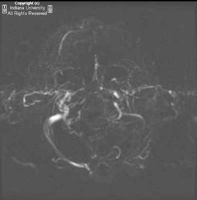
The head CT demonstrates diffuse cerebral edema, with findings of uncal herniation. Brain MRI showed multiple foci of restricted diffusion, including involvement of the right basal ganglia, thalami, and white matter. MRA demonstrates thrombosis of the left transverses sinus. MRA (arterial) of the brain suggested narrowing of the major intracranial vessels.

Differential Diagnosis:
- Viral encephalitis
- Carbon monoxide poisoning
- Leigh syndrome
- Infiltrating neoplasm

Fluorescent antibody and PCR were tested, which were positive for rabies. The child did not recover. Post mortem evaluation was consistent with rabies encephalitis.


Diagnosis: Rabies encephalitis, confirmed by laboratory evaluation and post mortem autopsy


Discussion

Rabies is caused by a RNA virus in the rhabdovirus family. The diagnosis of rabies encephalitis is clinical. Presenting symptoms may be non specific, but there is a typical presenting history of recent bat or other wild animal bit. The incubation period is between one and three months. According to the CDC, there have been only 6 documented cases of human survival from clinically evident rabies. Imaging is usually not performed because of the fatal and fulminate course

On imaging, the encephalitis form of human rabies demonstrates involvement of the brainstem, hippocampi, thalami, basal ganglia, and white matter. There is variable enhancement, and there is frequently restricted diffusion and diffuse cerebral edema. Intracranial arterial narrowing has been described in case reports.

Patients with carbon monoxide poisoning classically presents with abnormalities of the globus pallidi, with hypodensity on CT and hyper intensity of T2 weighted imaging. There is usually associated immediate neurological deficits, which persists over time.

Leigh syndrome is a mitochondrial disorder resulting in progressive neurologic deterioration. Adult presentation is unusual, with the majority of patients presenting by 2 years old. Imaging demonstrate bilateral hyper intensities involving the putamina and peri-aqueductal gray matter.

Diffusely infiltrating astrocytoma is usually unilateral and do not demonstrate restricted diffusion. There is variable enhancement.

giovedì 6 marzo 2008

Carotid Cavernous fistula (C-C fistula)


Findings

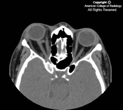
Figure 1: A non-contrast axial image through the upper orbits demonstrates an enlarged right superior opthalmic vein. Compare this vessel to the normal left superior opthalmic vein.
Figure 2: This image demonstrates proptosis, an enlarged medial rectus muscle, and a dilated superior opthalmic vein.
Figure 3: One slice below, we see enlarged medial and lateral rectus muscles.


Diagnosis: Carotid Cavernous fistula (C-C fistula)


A carotid cavernous fistula (C-C fistula) is a direct communication between the intracavernous portion of the carotid artery and the venous cavernous sinus. It is typically a result of trauma, either penetrating or non-penetrating, or a ruptured intracavernous carotid aneurysm. Other less common causes include Ehlers-Danlos and fibromuscular dysplasia due to intrinsic weakening of the vessel wall. This abnormal communication is most often found unilaterally. A patient with a C-C fistula usually presents with an eye motility disturbance, proptosis, an orbital bruit, and/or chemosis. Venous tributaries may connect the right and left cavernous sinuses, so that occasionally symptoms can be bilateral.

The pathophysiology of a C-C fistula begins with the release of arterial blood into the low-pressure cavernous sinus. The high arterial pressure decompresses into any venous structure that communicates with the cavernous sinus. Retrograde venous drainage leads to enlarged ophthalmic veins followed by venous congestion. The congestion causes an increase in the size of the extra-ocular muscles and orbital fat infiltration. This creates orbital mass effect and proptosis.

A CT examination demonstrates enlargement of the extra-ocular muscles, proptosis, and an enlarged superior opthalmic vein. A distended cavernous sinus can also be seen within the middle cranial fossa. MR imaging shows similar findings with the added ability to determine whether there is flow within opthalmic veins or if thrombosis has developed. MRI also allows for the detection of a carotid cavernous aneurysm if it is present. Catheter angiography of the external and internal carotid arteries and the vertebral arteries is necessary for accurate identification of the fistula; the lateral projection of the cranium is the most helpful. The angiographic appearance of a C-C fistula shows early, immediate, filling of the cavernous sinus and the dilated superior ophthalmic vein.

Although C-C fistulas can thrombose spontaneously, they are usually treated via an endovascular approach. The first option in treatment consists of using detachable coils or balloons, flow directed through the fistula into the cavernous sinus. Once within the sinus, the coils or balloons would tamponade the hole. If this doesn’t work, then another option would be to trap the fistula above and below its origin with balloons and/or surgical intervention.

Dural malformations involving the cavernous sinus can present with similar findings, but they differ from C-C fistulas in that they are not usually related to a traumatic event and the symptoms are not usually as severe.

mercoledì 5 marzo 2008

Diastematomyelia of cervical spine


Findings

T2 images demonstrate complete splitting of the cervical spinal cord into 2 complete hemicords without associated syrinx. The cord splits just caudal to the medulla and reunites distally at the upper thoracic spine. There are associated cervical segmentation anomalies, including intersegmental laminar and vertebral body fusion, without a dividing spur.

Differential Diagnosis:
- Diastematomyelia
- Myelomeningocele
- Syringomyelia
- Diplomyelia


Diagnosis: Diastematomyelia of cervical spine


Key points

Diastematomyelia usually presents in childhood with a female predilection. Adult presentation is rare, but when patients present older, there is a slight male predilection. Patients usually present with stable or progressive disability, scoliosis, and neurologic and muscular abnormalities. Often, Diastematomyelia is clinically indistinguishable from a tethered cord.
Treatment consists of surgical tethered cord release, spur resection, scoliosis correction, and dural repair.
Diastematomyelia occurs between T9 and S1 85% of the time.
Diastematomyelia often occurs when a fibrous osseous spur splits the cord sagittally into 2 hemicords that usually unite cranial and caudal to the cleft. In the patient presented above, the salient lesion was a lipoma, which was surgically resected.
85 % of Diastematomyelia patients have associated congenital spinal anomalies such as segmentation and fusion anomalies, intersegmental laminar fusion, dysraphism, tethered cord, syringohydromyelia, scoliosis, and Chiari II malformations. Intersegmental laminar fusion is virtually pathognomonic.
Meningocele manqué – Tethered cord due to nerve roots adhering to the dura.
Half of diastematomyelia patients present with cutaneous stigmata such as a "fawn's tail" hair patch at the level of the lesion.


Pang Classification

Type I Split cord malformation
- Separate dural sac
- Osseous spur
- More commonly symptomatic

Type II Split cord malformation
- Single dural sac
- No osseous spur, may see adherent fibrous bands tethering cord
- Usually asymptomatic unless associated with tethering or hydromyelia