Visualizzazione post con etichetta Ophtalmic. Mostra tutti i post

mercoledì 22 dicembre 2010

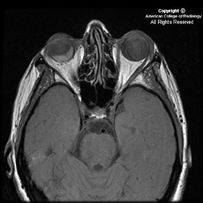
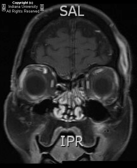
Intracranial pseudotumor (Tolosa-Hunt syndrome)


Findings

Increased CSF signal demonstrated by T2 hyper intensity within the right optic sheath. Nodular enhancement at the superior aspect of the right optic nerve at the orbital apex, which extends with prominent nodular enhancement posteriorly along the dural reflection of the right cavernous sinus. Asymmetric dilatation of the right superior ophthalmic vein.

Differential diagnosis:
- Meningitis
- Sarcoidosis
- En plaque meningioma
- Meningeal metastases
- Meningeal Non-Hodgkin's lymphoma
- Tolosa-Hunt syndrome


Diagnosis: Intracranial pseudotumor (Tolosa-Hunt syndrome)


Discussion

Intracranial pseudotumor (Tolosa-Hunt syndrome) is a part of a spectrum of "quasineoplastic" lesions that demonstrate orbital, intracranial, or pulmonary involvement, and include such other disorders as plasma cell granuloma and hypertrophic cranial pachymeningitis. It is a chronic granulomatous disease of unknown origin, which has been hypothesized to represent a low grade fibrosarcoma of inflammatory cells versus an autoimmune phenomenon. While Tolosa-Hunt syndrome is rare, orbital pseudotumor is the third most common ophthalmic disorder, and encompasses 5-8% of all orbital masses. Histologically, the lesions of Tolosa-Hunt demonstrate mixed lymphocytic and plasma cell infiltrate, with a variable degree of fibrosis. Lesions favor the cavernous sinus and basal meninges, although falcine and tentorial lesions have been described.

Patients with intracranial lesions are more frequently young adults, who present initially with chronic headaches or cranial nerve palsies. Patients with orbital involvement are more frequently middle-aged, and may present with painful proptosis and vision loss . Symptoms may be intractable, leading to severe disability. Untreated or unresponsive disease may progress to death. First-line treatment is invariably high-dose steroids, with radiotherapy or surgical resection reserved for patients with incomplete response to steroids .


Radiologic Overview of the diagnosis

The imaging hallmarks of Tolosa-Hunt syndrome are characterized by an enhancing, infiltrating meningeal mass, which favors the cavernous sinus or basal meninges, although falcine and tentorial involvement has been described . Focal meningeal thickening may range from just a few millimeters to a greater than 2 cm rind. With intracranial pseudotumor, orbital involvement is spared more than 90% of the time. Tolosa-Hunt remains a diagnosis of exclusion, once meningitis, en plaque meningioma, and meningeal metastases are ruled out.

The imaging modality of choice for imaging patients with Tolosa-Hunt remains MRI, although useful information may be gleaned from other modalities. On non-contrast enhanced CT (NECT), there are no specific findings to suggest the diagnosis; however, this modality may be of some value in differentiating the lesion from en plaque meningioma. On contrast enhanced CT (CECT), salient imaging findings include enhancing, thickened meninges or a curvilinear appearance of a single meningeal region. As aforementioned, MRI remains the primary modality for diagnosis of Tolosa-Hunt syndrome, and each sequence may provide critical information required to make the diagnosis. On T1WI, one may find focal thickening of the meninges that is isointense to gray matter. On T2WI, lesions are characterized as iso- to hypo intense regions of focal meningeal thickening, which may be more hypo intense as they become more fibrotic. FLAIR is of little help in making the diagnosis, but it is unlikely to demonstrate focal brain edema underlying the lesion . Contrast enhanced T1WI is the single most valuable sequence for evaluation of Tolosa-Hunt syndrome, and is characterized by diffusely enhancing region of meningeal thickening, which may range from a few millimeters to greater than 2 cm in some cases. Diffuse boney infiltrates may be appreciated on fat saturated contrast enhanced T1 sequence. While angiography is not considered a primary modality, severe disease may result in carotid artery narrowing, thus MRA may be a useful adjunct in the appropriate clinical setting.

The appropriate differential diagnosis of Tolosa-Hunt syndrome includes meningitis, sarcoidosis, en plaque meningioma, meningeal metastases, and meningeal Non-Hodgkin's lymphoma.

venerdì 19 novembre 2010

Malignant melanoma of the uvea


Findings

Thin section axial fat-suppressed fast spin-echo T2-weighted imaging
Figure 1 and Figure 2 show a V-shaped retinal detachment pointing toward the optic nerve. There is increased T1 signal posterior to the detachment suggesting hemorrhage.

Thin section axial fat-suppressed T1-weighted imaging
Figure 3 and Figure 4 show a mass arising from the nasal aspect of the right ocular globe. There is a collar button configuration to this mass, which assumes the typical configuration strongly suggesting elevation of Bruch’s membrane. The mass is near isointense to muscle on precontrast T1 and T2 weighted images. This mass extends towards the vitreous but no extension beyond the sclera is identified.

Postcontrast fat-suppressed T1-weighted imaging
Figure 5 and Figure 6: The mass is near isointense to muscle on pre-contrast T1 and T2 weighted images and demonstrates prominent contrast enhancement.


Diagnosis: Malignant melanoma of the uvea


Uveal melanoma is the most frequent primary intraocular malignant tumor in adults. It is rare in children. Its importance is that it is the main intraocular disease that can be fatal in adults.
Uveal melanoma can erupt through Bruch's Membrane. When this occurs, they develop a characteristic collar button configuration that extends through the vitreous chamber.
Uveal melanomas have differing MR characteristics, depending on the amount of melanin, which has paramagnetic properties. Melanomas containing a lot of melanin will demonstrate T1 hyperintensity, and markedly decreased T2 signal. However, in amelanotic or slightly melanotic melanomas, the typical MR pattern is isointense on T1, and slightly hypointense on T2.

Uveal melanoma is the most frequent form of intraocular malignancy in adulthood (4). As the uvea is the most vascular region of the globe, it is a common site for primary and metastatic neoplasm. Uveal melanomas comprise 70% of malignant intraocular tumors. It affects approximately 5-7 out of 1,000,000 people (3). Uveal melanoma can occur in any of the three subdivisions of the uvea: the iris, ciliary body, and choroid.

Uveal melanomas start with a flat growth profile along the choroid. With progression, they become elevated, and frequently extend through Bruch's membrane, where they can track into the subretinal space. With spread through Bruch's membrane, the melanoma can have a "mushroom shape" or "collar button" appearance that extends through the vitreous chamber.

Uveal melanomas typically appear as a solid, well-defined mass on magnetic resonance imaging. Melanin is paramagnetic, so in melanomas containing a lot of melanin, there is increased T1 signal with markedly decreased T2 signal. This signal intensity pattern is pathognomonic for uveal melanoma, as there are no other intraocular lesions with this appearance. In low-melanin or amelanotic melanomas, Magnetic Resonance imaging is less specific, but typically shows isointense signal on T1-weight images and slightly hypointense signal on T2-weighted images. Uveal melanomas typically have moderate to strong contrast enhancement following administration of gadolinium.

B-mode ultrasound typically shows a rounded, hypoechoic, highly vascular lesion. Retinal elevation and vitreous hemorrhage can also be seen, as these are complications of uveal melanomas. Uveal melanomas on unenhanced computed tomography appear sharply marginated, hyperattenuating, and elevated.

Uveal melanomas also have a propensity to metastasize hematogenously, and do so most frequently to the liver. Uveal melanoma is the most common fatal intraocular disease in the adult population.

Optimal treatment for uveal melanomas is controversial and clinical trials are ongoing. Large melanomas, typically greater than 10-mm in thickness, are usually managed with enucleation. For medium sized melanomas, 3-mm to 10-mm thick, plaque brachytherapy and external-beam radiation therapy have been accepted as alternatives to enucleation. For small lesions, less than 3-mm, routine monitoring with ultrasound is recommended as these may represent benign choroid nevi. These small lesions may also be biopsied, with a positive result placing the small melanomas into the medium melanoma treatment category.

Prognosis is dependent on many factors. Increasing tumor pigmentation is associated with a less favorable prognosis. Additionally, increasing size, infiltration through Bruch's membrane, and retinal detachment are all associated with a poorer prognosis. In metastatic disease to the liver, the mean survival has been reported to be nine months.

lunedì 1 novembre 2010

Retinoblastoma with recurrence following enucleation


Findings

Figure 1: Contrast enhanced CT scan shows a heterogeneously enhancing intra-ocular mass in the right eye with calcification.
Figure 2: Contrast enhanced CT image shows enlargement of the right optic nerve suggestive of orbital recurrence following enucleation. Note the presence of a prosthetic globe.


Diagnosis: Retinoblastoma with recurrence following enucleation


The differential diagnosis of leukocoria in a child includes retinoblastoma, Coat’s disease, toxocariasis, retinopathy of prematurity, optic nerve drusen, persistent hyperplastic primary vitreous and phthisis bulbi. With a normal sized globe and intra-ocular calcifications the differential narrows down to retinoblastoma, Coat’s disease and toxocariasis with retinoblastoma being the most common.

Most patients with unilateral, non-metastatic retinoblastoma can be cured with enucleation alone. In patients with histologic risk factors, adjuvant chemotherapy is recommended, with the addition of orbital radiation for patients with trans-scleral involvement or tumor present at the level of the cut end of the optic nerve. Patients with metastases require intensive chemotherapy and consolidation with autologous hematopoietic stem cell rescue.

Recurrent retinoblastoma or a second primary neoplasm may be observed during follow-up. Optic nerve invasion, scleral invasion and choriodal invasion have been regarded as risk factors for recurrent disease. In general recurrent tumors have a short latent period as compared to second primary neoplasms; however this may not always be true. Recurrent retinoblastoma after enucleation can occur in the contralateral eye (type A), in the ipsilateral orbit (type B) or extend from the orbit into the brain (type C)

The most common presentation of orbital recurrence is a clinical complaint such as eyelid swelling or chemosis, suggesting that clinicians should not attribute periocular or orbital symptoms during the first year after enucleation as a sign of a benign conjunctivitis or preseptal cellulitis. A problem with the ocular prosthesis is also a frequent mode of presentation in these patients, and extrusion or displacement of a previously satisfactory prosthesis should be considered a suspicious sign for possible tumour recurrence.

Calcification maybe seen within the recurrent tumors on unenhanced CT. Most recurrent tumors appear iso to hypointense to normal temporal muscle on T1 weighted images and show moderately high signal on T2 weighted images. Contrast enhanced scans, CT or MRI, show heterogeneous enhancement of the tumor.

Cases with recurrence following enucleation should be evaluated for distant metastasis. Treatment consists of systemic chemotherapy combined with other modalities like orbital radiotherapy and stem cell rescue, depending upon the extent of metastatic disease.

mercoledì 13 ottobre 2010

Intraorbital Lymphatic Malformation


Findings

Figure 1: There is a multilobulated mass lesion in the retroorbital region with a fluid- fluid level.
Figure 3: There is an intraconal multilobulated mass with a fluid-fluid level and mild right globe proptosis.


Diagnosis: Intraorbital Lymphatic Malformation


Vascular lesions account for 5-20% of all orbital masses, and the two most common orbital vascular lesions are venous malformations (formerly known as cavernous hemangiomas) and lymphatic malformations (LM) ( formerly known as lymphangiomas). LMs are relatively uncommon in the pediatric population and account for only 4% of all childhood orbital masses. LMs are benign and most frequently found in the head and neck. Intraorbital LMs can arise in any orbital space, but are most commonly intraconal with frequent extraconal and preseptal expansion. Histologically, a vascular malformation can contain venous and lymphatic components, hence the name lymphaticovenous malformation. Of note, 70% of orbital lymphaticovenous malformations are associated with ipsilateral, noncontiguous, intracranial vascular abnormalities.

Intraorbital venous-lymphatic malformations are present at birth, but tend not to be discovered clinically until early childhood when they enlarge as a result of either intralesional hemorrhage or lymphoid hyperplasia and result in acute proptosis. Approximately one-half of all patients with orbital LMs also complain of limited ocular mobility. Additionally, conjunctival, facial, or oral vesicles may also be observed.

Radiologic imaging of intraorbital LMs demonstrates unencapsulated, irregular, lobulated, and multicompartmental masses. These lesions can have cystic as well as more solid components. The cystic elements of these masses commonly exhibit fluid-fluid levels as a result of intralesional hemorrhage. Additionally, LMs are frequently both pre- and postseptal and intra- and extraconal. They often display orbital expansion with irregular margins that traverse tissue planes. Ultrasound images of LMs demonstrate heterogeneous, ill-defined lesions with anechoic cystic portions and extraconal extension. On CT, these masses exhibit ill-defined borders, irregular attenuations, and variable enhancement with peripheral rim enhancement in cystic regions. Additionally, calcified phleboliths can be seen on CT in venous portions of these lesions. MR imaging is the preferred imaging modality to evaluate the location, vascular components and evolving blood products of venous-lymphatic malformations. LMs demonstrate iso- to slightly high signal intensities on T1-weighted images and very high signal intensities on T2-weighted images. MR imaging also allows for the simultaneous evaluation of the brain in an effort to detect any associated intracranial vascular anomalies.

The differential diagnosis for pediatric orbital tumors can be divided into osseous and non-osseous lesions. Pediatric osseous lesions of the orbit include dermoid inclusion cysts, which are most common, fibrous dysplasia, juvenile ossifying fibroma, osteosarcoma, Langerhans cell histocytosis, granulocytic sarcoma and neuroblastoma bone metastases. Non-osseous lesions of the pediatric orbit include most commonly rhabdomyosarcoma, but additionally infantile fibromatosis, infantile hemangioma, and LM.

Orbital lymphaticovenous malformations are histologically benign, but can demonstrate aggressive behavior, such as vision loss, as they expand. The treatment of such lesions is focused on ameliorating pain, alleviating optic nerve compression, maintaining ocular alignment and improving cosmetic appearance. Surgical resection is the preferred treatment for these lesions, and while complete removal can often be achieved with well-demarcated extraconal lesions, more diffuse intraconal lesions tend to be treated with subtotal resection.

mercoledì 1 settembre 2010

Bilateral ectopia lentis


Findings

Figure 1: Axial noncontrast CT of the head at the level of the orbits demonstrates posterior dislocation of both lenses, which now rest dependently in the vitreous. The etiology in this particular patient was repetitive trauma from serial falls.


Diagnosis: Bilateral ectopia lentis


The crystalline lens of the eye is designed to refract the light entering the iris and project/focus it onto the retina. The lens itself contains no vasculature, nerves, or connective tissue. It sits behind the iris and the front of the lens is in contact with the aqueous fluid of the anterior chamber while the posterior surface of the lens is in contact with the vitreous. The lens is held in place by zonular fibers, otherwise known as suspensory ligaments. These fibers connect to the cilliary body around the circumference of the lens.

Subluxation (partial dislocation) or luxation (complete dislocation) of the crystalline lens, otherwise known as ectopia lentis, is caused by dysfunction or disruption of these zonular fibers. Trauma is the most common cause of this disorder. The absence of a traumatic history should prompt consideration of hereditary causes of zonular fiber dysfunction; predisposing conditions include Marfan syndrome, homocystinurea, tertiary syphilis, and Weil-Marchesani syndrome.

Patients will complain of monocular diplopia, markedly decreased visual acuity in the affected eye(s), and/or poor near vision.

Treatment is determined by lens position, with anterior chamber dislocation often being a surgical emergency. As the aqueous humor of the eye flows in the anterior chamber, around the iris from the cilliary body to the canal of Schlemm, this route can become acutely obstructed with anterior dislocation leading to acute glaucoma. The cornea and iris are also at risk for damage. Posterior dislocation may be treated conservatively depending on lens position, but may also lead to uveitis or glaucoma in some cases.

venerdì 6 agosto 2010

Sinonasal and orbital lymphoma


Findings

Figure 1: Coronal image from CECT shows enhancing mass involving the maxillary and ethmoid sinuses and left orbit.
Figure 2: Coronal image from CECT shows mass in maxillary sinus causing dehiscence of floor of left orbit.
Figure 3: Axial image from CECT shows enhancing mass in ethmoid sinuses and intraconal portion of left orbit causing proptosis.


Diagnosis: Sinonasal and orbital lymphoma (Diffuse Large B-cell Lymphoma)


Malignant neoplasms of the nasal cavity and paranasal sinuses occur primarily in the 5th to 6th decade. Exposure to industrial fumes, leather tanning and even wood dust have been implicated in the carcinogenesis of certain malignant sinonasal tumors. Interestingly cigarette smoking and heavy alcohol consumption have not been linked to sinonasal cancers despite there strong association with other head and neck cancers.

Lymphomas of the sinonasal tract are uncommon neoplasms that can be clinically and radiologically difficult to distinguish from destructive non-malignant processes such as aggressive sinusitis or benign neoplasms. Primary sinonasal lymphoma is rare in western populations, but is more common among Chinese and Japanese populations. Approximately 2000 cases of sinonasal cancer are diagnosed each year in the United States. Lymphoma accounts for less than 5% of all sinonasal cancers and less than 1% of all head and neck cancers.

Low grade lymphomas usually present with a nasal cavity or paranasal sinus mass associated with obstructive symptoms. High grade lymphomas are more likely to present with aggressive signs and symptoms including nonhealing ulcer, cranial nerve manifestations, facial swelling, epistaxis, or pain. Of note, the high grade B-cell lymphomas tend to present with soft tissue or osseous destruction, particularly of the orbit with associated proptosis, whereas T-cell lymphomas are associated with nasal septal perforation and/or destruction.

In Asian populations Natural Killer or T-cell (NK/T-cell) immunophenotypes are more common then B-cell where-as in western populations, there seems to be a slight B-cell predominance. Prognostically NK/T-cell lineage lymphomas have a worse outcome then B-cell. The reason of this discrepancy is not clear but may be related to NK/T-cell frequent expression of an energy-dependent efflux pump capable of transporting chemotherapeutic drugs out of the cell.

Computed tomography (CT) and magnetic resonance imaging (MRI) play complementary roles in determining the full extent of a sinonasal neoplasm. CT is useful to detect bone erosion but can be limited to differentiate the extent of the tumor particularly as the mass approaches the periobita. MRI provides excellent delineation of tumor from surrounding inflammatory tissue and secretions within the sinuses which often accompany obstructive masses. Most sinonasal tumors are highly cellular and give intermediate signal intensity on both T1 and T2 weighted imaging and demonstrate diffuse enhancement.

lunedì 26 luglio 2010

Chronic progressive external ophthalmoplegia


Findings

The extraocular muscles are atrophic and to some degree show fatty replacement. No abnormal enhancement is present. The globes are intact bilaterally. The intraorbital fat appears slightly increased. There is bilateral orbital proptosis with both globes anterior to the interzygomatic line.


Diagnosis: Chronic progressive external ophthalmoplegia


Key points

Chronic progressive external ophthalmoplegia (CPEO) is characterized by slowly progressive paralysis of the extraocular muscles.
Mitochondrial myopathy usually associated with skeletal muscle weakness.
Presents with bilateral, symmetrical, progressive ptosis, followed by ophthalmoparesis months to years later.

Kearns-Sayre syndrome: Related mitochondrial myopathy with CPEO, onset before age 20 years, pigmentary retinopathy, and at least one of the following: cardiac conduction defects, CSF protein of greater than 100 mg/dL, and/or cerebellar syndrome.
KSS can include mental retardation, hearing loss, seizures, short stature, delayed puberty, and various endocrine disorders.
Frequency: Rare. Males=Females.

Imaging studies: MRI, CT, and ultrasound may show thin, symmetrical extraocular muscles in CPEO, in contrast to enlarged extraocular muscles sometimes seen with Graves disease.
Those with KSS and CPEO display a wide spectrum of MRI findings, including normal brain, diffuse atrophy, and T2 prolongation in subcortical cerebral white matter, cerebellar white matter, globi pallidi, thalami, and substantia nigra.
Diagnosis: Muscle biopsy is definitive test but PCR also shown to be conclusive.

lunedì 7 giugno 2010

Orbital Dermoid


Findings

Figure 1 and Figure 2: Ovoid well-circumscribed ventromedial left orbital extraconal mass resulting in mass effect on the intraorbital contents displacing the globe laterally, demonstrating nonaggressive features. Internal areas of fat attenuation.

Figure 3and Figure 4: Ovoid well-circumscribed mass centered in the post-septal, extraconal left orbit which exerts mass effect on the left globe, displacing it laterally. Signal characteristics of the lesion are most consistent with an intraorbital dermoid, including fat signal layering within the nondependent portion of this predominantly cystic lesion. This fat floats on underlying proteinacous cystic material.


Diagnosis: Orbital Dermoid


Patients with this diagnosis generally complain of an orbital mass, which may be visible on physical examination. Growth of these lesions is generally slow. In adults, dermoids may become symptomatic for the first time and grow considerably over a year. Based on this fact, some authors have concluded that these lesions may be dormant for many years and/or have intermittent growth, such as in our case example.

Dermoids are the most common benign congenital lesion of the orbit, accounting for 1-2% of all orbital masses. Dermoid and epidermoid cysts are examples of choristomas, tumors that originate from aberrant primordial tissue. These tumors contain normal-appearing tissue in an abnormal location. As two suture lines of the skull close during embryonic development, dermal or epidermal elements are pinched off and form cysts, which are adjacent to the suture line. Approximately 50% of these tumors that involve the head are found in or adjacent to the orbit. They arise most often in the superolateral portion of the orbit at the frontozygomatic suture. They can also arise inferiorly, posteriorly, or medially (as in our case example). These lesions are extraconal and displace the globe. If the displacement is great, interference with vision by compression of the optic nerve may result or ocular motility may be disturbed, typically resulting in diplopia.

On MR the diagnosis is usually clinched with high signal on T1WI in the region which suppresses with fat saturation techniques. Fat may be seen floating in cystic fluid on T1WI.

These tumors are most often noted in young children; however, they may appear or grow at any age. In adults, they are more likely to displace the globe, possibly growing or eroding their way into adjacent

giovedì 13 maggio 2010

Poorly differentiated orbital malignant lymphoma


Findigs:

Figure 1: Axial CT through the orbital floors at the level of the inferior orbital foramen demonstrates an erosive mass, containing calcifications, with expansion through the floor of the orbit and into the maxillary sinus.
Figure 2: Contrast enhanced coronal CT image demonstrates a heterogeneously enhancing, soft tissue mass involving the floor of the orbit, with bony invasion.


Diagnosis: Poorly differentiated orbital malignant lymphoma (Non-Hodgkin's type) (biopsy proven)


In the adult population the differential diagnosis of malignant orbital tumors differs from that of children. Whereas, carcinoma is the frequently enountered in adults (often from contiguous spread from the paranasal sinus, etc), metastatic neuroblastoma is the more frequently encountered malignant neoplasm in children.

Neoplasms of the paranasal sinuses are uncommon, but frequently extend to involve the orbit when they do occur. Benign tumors tend to push the periorbital tissues aside, while malignant lesions tend to invade the periosteum.

Evaluation of the paranasal sinus mass is best done utilizing multidetector CT, because of its ability to detect bony destruction and possible intracranial extension of disease. CT usually demonstrates a homogeneous mass with well-defined borders that demonstrates heterogeneous enhancement.

Lymphoid tumors are one of the most common orbital tumors despite the orbit not containing lymph nodes or a well defined lymphatic vasculature. Ocular malignant lymphomas are uncommon. The incidence ranges from 1-4%. Involvement of the orbit as a presenting sign is rare and few such cases are seen in the literature. Although most orbital lymphomas are localized to the orbit at diagnosis, many patients will develop systemic lymphoma over time. Orbital lymphoma is an adult disease, usually presenting in patients between the age of 50-70 years. The course is usually of an anterior mass that causes painless proptosis over several weeks to months.

Radiation therapy is the mainstay of treatment since surgical resection plays a limited role. Response rate and prognosis depends upon cell type, the extent of disease and whether the orbital disease represents the primary site or the site of spread of disease from an extra-ocular primary. Five year survival rates range from 54-78%.

venerdì 26 febbraio 2010

Sarcoidosis involving the lacrimal glands


Findings

Axial (Figure 1) and coronal (Figure 2) non contrast CT images, as well as an axial T2 MRI image (Figure 3), demonstrate symmetrically enlarged lacrimal glands which protrude anterior to the lacrimal fossa bony contours. Homogeneous enhancement is identified on the selected contrast enhanced axial CT image (Figure 4). There is no evidence of adjacent bony orbit invasion. An AP chest radiograph (Figure 6) demonstrates predominantly right hilar lymphadenopathy with “eggshell” calcification and bilateral reticulonodular opacities within the lung parenchyma.


Diagnosis: Sarcoidosis involving the lacrimal glands


20 to 25 percent of patients with systemic sarcoidosis develop ophthalmic manifestations usually between the third and fifth decades. The most common finding is inflammation of the uveal tract. Less commonly, patients will present with inflammation of the optic nerve or orbital involvement including pseudotumor like intraorbital masses, extraocular muscle enlargement, or lacrimal gland infiltration and hypertrophy. Isolated orbital disease is uncommon and usually limited to the lacrimal glands.

Lacrimal gland involvement occurs in approximately 15-28% of patients; usually as painless bilateral gland swelling evident on physical examination. In many cases, lacrimal gland involvement may occur long before lung and other organs are affected, thereby aiding in the early diagnosis of systemic sarcoidosis.

Imaging studies such as orbital CT and MRI are an integral part of early diagnosis. CT findings include symmetric enlargement of the lacrimal glands with diffuse homogeneous post contrast enhancement. There may be associated medial displacement of the optic globes as well as proptosis. Pertinent negative findings include the absence of adjacent bony orbital invasion and destruction. MRI is optimal to evaluate for additional orbital involvement such as optic nerve infiltration which manifests as a thickened and enhancing intraorbital nerve. An MRI of the brain is also recommended to evaluate for extension into the intracranial optic pathways and to exclude findings of coexisting neurosarcoidosis.

While diagnostic imaging tests may reveal findings highly suggestive of lacrimal gland sarcoidosis; definitive diagnosis requires biopsy of the glandular tissue and histopathologic assessment. Noncaseating granulomas characterized by clustered epithelial cells, central giant cells and abundant surrounding lymphocytes are characteristic.

The mainstay of therapy is high dose systemic steroids, usually oral prednisolone for approximately two weeks, followed by gradual tapering after the inflammation appears controlled. Some patients may require maintenance doses for several weeks to months.

lunedì 15 febbraio 2010

Bilateral orbital metastatic scirrhous carcinoma of the breast


Findings

Figure 1: In the right orbit region there is a retroglobar soft tissue mass with no associated proptosis. Retraction of the globe is appreciated.
Figure 2: Axial T1 post contrast with fat saturation. There is heterogeneously enhancing infiltrative appearance to the bilateral retrobulbar compartments, right greater than left. There is also associated infiltration and fullness of the orbital fat.


Diagnosis: Bilateral orbital metastatic scirrhous carcinoma of the breast


Orbital metastasis is a significant cause of orbital disease in adults and accounts for 3-10% of all orbital tumors. While embryonal tumors, neuroblastomas, Ewing’s sarcomas and Wilms tumors are responsible for most pediatric metastatic orbital tumors, orbital metastases in the adult population are predominantly from breast, lung, and prostate carcinomas. In the female population, the most common primary tumor metastasizing to the orbit is adenocarcinoma of the breast. While only 8% of breast cancers demonstrate symptomatic metastatic disease at initial presentation, 12-31% of cases exhibit orbital metastases as the earliest indication of metastatic spread. Furthermore, when orbital metastases are detected they are frequently associated with pervasive metastatic disease.

Orbital metastatic lesions typically present in the 7th decade of life. They tend to arise in the choroid, retrobulbar soft tissues and bony orbit. While it is uncommon for orbital metastases to present bilaterally, breast carcinoma is the most likely malignancy to be associated with bilateral orbital metastases. Clinically, orbital metastases demonstrate acute proptosis, external opthalmoplegia, visual deficits, diplopia, scotomata, and orbital pain. Additionally, scirrhous adenocarcinoma of the breast often presents with enopthalmos, which has been hypothesized to result from the orbital fibrosis generated by the tumor.

Radiologic imaging of orbital metastases of scirrhous carcinoma of the breast typically demonstrates diffusely infiltrating retrobulbar masses with ill-defined margins. On CT, metastases to the choroid or retina are typically seen in the posterior globe with frequent expansion into the retrobulbar space. These metastases also have the potential to cause secondary retinal detachment and hemorrhage. Metastases to the extraocular muscles, optic nerve, or lacrimal gland are associated with an increase in the structure’s size, irregular borders and slight contrast enhancement. Additionally, MR imaging may also be used to evaluate orbital metastatic lesions. An added benefit associated with MRI is the ability to simultaneously detect concurrent silent brain lesions, which are not infrequently found along with orbital breast metastases.

The differential diagnosis for an orbital lesion in an adult is diverse and includes vascular tumors (cavernous hemangiomas), meningiomas, lymphomas, cysts, lacrimal gland tumors, inflammatory masses (pseudotumors), and carcinomas (primary and metastatic).

Fine-needle aspiration biopsy is often utilized to confirm the diagnosis of orbital metastases. In the case of metastatic scirrhous carcinoma of the breast such biopsies tend to be more challenging as a result of the fibrous stroma that characteristically encases these malignancies. Treatment of orbital metastases is primarily local radiotherapy; however, chemotherapy is often required with systemic disease. Following the discovery of distant metastases, there is a 2 year survival associated with metastatic breast cancer.

martedì 12 gennaio 2010

Optic disc coloboma


Findings

A posterior protrusion of vitreous is seen in the left globe, at the insertion of the optic nerve head. There is no hemorrhage, retrobulbar colobomatous cyst, or optic nerve atrophy. The right globe is normal.

Differential diagnosis:
- Coloboma (chorioretinal or optic disc)
- Peripapillary staphyloma
- Morning glory disc anomaly (MGDA)
- Buphthalmos (ox eye)


Diagnosis: Optic disc coloboma


Key points

Coloboma (Greek koloboun, "to mutilate") is the result of incomplete closure of the choroidal fissure.
Funnel or cone-shaped protrusion from the posterior aspect of the globe, often involving the optic nerve head insertion.
Two general types: Optic disc coloboma (ODC = excavated/everted region involves the posterior globe at the optic disc insertion only). Chorioretinal colobomas are more broad-based, extending beyond the margins of the optic disc.
May be associated with large retrobulbar colobomatous cysts (apparently encapsulated outpouchings of vitreous) which may be larger than the actual globe, may cause significant proptosis, or even globe atrophy / microphthalmia.
Majority of colobomas are sporadic and unilateral.
Associated with midline craniocerebrofacial clefting, choanal atresia, basal (particularly sphenoidal) encephalocele, corpus callosal agenesis, olfactory hypoplasia, cardiac anomalies, retardation, genital hypoplasia, and ear anomalies.

Differential diagnosis:
- Peripapillary staphyloma – more diffuse / broad based than coloboma, must feature posterior uveoscleral thinning and lack enhancement.
- Morning glory disc anomaly – fundoscopic description of ODC. Funnel or square shaped posterior defect at the optic nerve head.
- Buphthalmos ("ox eye") – 2/2 congenital glaucoma. Assoc. with Marfan's, NF-1, Sturge-Weber. Entire globe enlarged.

mercoledì 28 ottobre 2009

Carotid cavernous fistula


Diagnosis: Carotid cavernous fistula


Discussion

Carotid Cavernous fistula (CCF) is a direct communication between the intracavernous portion of the carotid artery and the venous cavernous sinus. Usually is ipsilateral.
Most often results from significant trauma, penetrating or nonpenetrating.
Spontaneous rupture happens in the elderly.

Spontaneous rupture of CCF also associated with:
- Osteogenesis imperfecta
- Ehlers-Danlos syndrome
- Psuedoxanthoma elastica

Clinical presentation: acute onset pulsating exophthalmos, orbital bruit, dilated conjunctival vessels, and glaucoma. No pain whereas psuedotumor is almost always associated with pain.


Radiology

Unilateral diffuse enlargement of extra ocular muscles.
Proptosis.
Dilation of Superior ophthalmic vein (SOV) and venous structures within the carotid cavernous sinus due to backpressure. Our case was unusual in that the venous engorgement was on the opposite side of the CCF.
Irregularity of SOV may represent thrombus (not seen in this case).
Bowed convexity to the Cavernous sinus usually unilateral (our case demonstrated right cavernous sinus bowing due to the venous engorgement from the opposite carotid-very atypical)
US: reversal of flow in SOV.
Carotid Angio: filling of ophthalmic veins due to decompression of arterial flow with retrograde filling of orbital veins.
Repair: important to determine the extent of contralateral flow in the cavernous sinus without the CCF if carotid must be sacrificed. Glue embolization of the SOV and coiling are the primary treatments.
Diagnosis first by CT orbit findings then proceed to conventional angiogram for diagnosis and treatment.
Treatment: glue embolization and coiling. Approach varies but may go through the SOV from the internal jugular or direct approach by interventional neuroradiologist or neurosurgeon through the SOV as in our case.


Pearls

With carotid cavernous fistula look for enlarged superior opthalmic vein.(SOV) in addition to unilateral rectus enlargement and proptosis.
Need cerebral angiogram to definitively diagnose-may not see enlarged carotid cavernous sinus on CT.
SOV lives between the superior rectus and the optic nerve.
Treatment of CCF is with glue embolization and coiling

giovedì 15 ottobre 2009

CHARGE syndrome


Findings

Posterior orbital globe defects of bilateral colobomas (Figure 1).
Bilateral choanal atresia, predominantly osseous component (Figure 2). Notice the bilateral air-fluid levels within the nasal cavity, which is classic for choanal atresia as fluid cannot drain posteriorly into the nasopharynx. There is deformity of both pinnae (Figure 3 and Figure 4).
Deformities of the semicircular canals (Figure 5).


Diagnosis: CHARGE syndrome

CHARGE is an acronym for the major clinical features of the syndrome (ocular Coloboma (75-90%), Heart defects (50-85%), Atresia of nasal choanae (35-65%), Retardation of growth/development, Genital anomalies (50-70% with male predominance), Ear anomalies (>90%). Additional associated features include facial dysmorphism, anosmia, auditory and vestibular anomalies, hypothalamo-hypophyseal dysfunction, and urinary tract anomalies. CHARGE syndrome occurs sporadically with an estimated prevalence of 1:10,000. Many patients with CHARGE syndrome have been genetically linked to mutations in the CHD7 gene, which encodes a protein within the chromodomain helicase DNA-binding (CHD) family of proteins responsible for gene expression regulation via chromatin remodeling. The ubiquitous expression of CHD7 protein helps explain the pleiotropic effects of patients with CHARGE syndrome.

Diagnosis of CHARGE syndrome initially required 4 of the cardinal 6 signs described by its acronym, with at least one sign being choanal atresia or coloboma. The diagnostic criteria were updated by Blake et al in 1998 to include brainstem anomalies, facial dysmorphism, and characteristic ear anomalies. More recently, and relying on the clinical triad of Coloboma-Choanal Atresia-semicircular Canal Anomalies, Verloes has proposed dividing the syndrome into typical, partial/incomplete, and atypical CHARGE syndromes based on 3 major signs (ocular coloboma, choanal atresia, hypoplastic semi-circular canals) and 5 minor signs (rhombencephalic dysfunction, hypothalamo-hypophyseal dysfunction, abnormal middle/external ear, mediastinal organ malformation, mental retardation).

Bilateral choanal atresia is usually osseous but may be membraneous and is usually detected in the newborn since neonates are obligate nose-breathers during feeding. Treatment is surgical with a transnasal or transpalatal approach to open the choanae. Ocular colobomas are often small and may only be detected via a funduscope. Vision is usually not impaired. Semicircular canal aplasia/dysplasia results in loss of nystagmus response to auditory caloric stimuli.