venerdì 26 febbraio 2010

Sarcoidosis involving the lacrimal glands


Findings

Axial (Figure 1) and coronal (Figure 2) non contrast CT images, as well as an axial T2 MRI image (Figure 3), demonstrate symmetrically enlarged lacrimal glands which protrude anterior to the lacrimal fossa bony contours. Homogeneous enhancement is identified on the selected contrast enhanced axial CT image (Figure 4). There is no evidence of adjacent bony orbit invasion. An AP chest radiograph (Figure 6) demonstrates predominantly right hilar lymphadenopathy with “eggshell” calcification and bilateral reticulonodular opacities within the lung parenchyma.


Diagnosis: Sarcoidosis involving the lacrimal glands


20 to 25 percent of patients with systemic sarcoidosis develop ophthalmic manifestations usually between the third and fifth decades. The most common finding is inflammation of the uveal tract. Less commonly, patients will present with inflammation of the optic nerve or orbital involvement including pseudotumor like intraorbital masses, extraocular muscle enlargement, or lacrimal gland infiltration and hypertrophy. Isolated orbital disease is uncommon and usually limited to the lacrimal glands.

Lacrimal gland involvement occurs in approximately 15-28% of patients; usually as painless bilateral gland swelling evident on physical examination. In many cases, lacrimal gland involvement may occur long before lung and other organs are affected, thereby aiding in the early diagnosis of systemic sarcoidosis.

Imaging studies such as orbital CT and MRI are an integral part of early diagnosis. CT findings include symmetric enlargement of the lacrimal glands with diffuse homogeneous post contrast enhancement. There may be associated medial displacement of the optic globes as well as proptosis. Pertinent negative findings include the absence of adjacent bony orbital invasion and destruction. MRI is optimal to evaluate for additional orbital involvement such as optic nerve infiltration which manifests as a thickened and enhancing intraorbital nerve. An MRI of the brain is also recommended to evaluate for extension into the intracranial optic pathways and to exclude findings of coexisting neurosarcoidosis.

While diagnostic imaging tests may reveal findings highly suggestive of lacrimal gland sarcoidosis; definitive diagnosis requires biopsy of the glandular tissue and histopathologic assessment. Noncaseating granulomas characterized by clustered epithelial cells, central giant cells and abundant surrounding lymphocytes are characteristic.

The mainstay of therapy is high dose systemic steroids, usually oral prednisolone for approximately two weeks, followed by gradual tapering after the inflammation appears controlled. Some patients may require maintenance doses for several weeks to months.

giovedì 25 febbraio 2010

Spinal meningioma


Findings

There is a contrast enhancing extramedullary, intradural lesion at T6.

Differential diagnosis:
- Meningioma
- Schwannoma
- Drop metastases
- Arachnoid cyst


Diagnosis: Spinal meningioma


Key points

Meningiomas are dural based, benign, slow growing tumors. They are usually solitary lesions and are 8 times more likely to occur in the brain than the spinal cord. When in the spinal cord, the thoracic spine is affected 80% of the time. On pathology, meningiomas are usually firm, round and well demarcated. Meningiomas have a peak incidence in the 5th and 6th decades of life. Presenting symptoms are usually due to mass effect the tumor has on adjacent neural tissue and can include pain, motor or sensory deficits.

Meningiomas are usually WHO grade I tumors (95%) and treated with surgical excision. In this case, the patient presented with back pain. She subsequently underwent resection of her tumor without recurrence.


Radiology

Meningiomas are well circumscribed, strongly enhancing lesions. On non-enhanced CT, meningiomas are difficult to recognize and usually isodense to mildly hyper dense when compared to surrounding brain parenchyma. With contrast, meningiomas enhance homogeneously. On T1 and T2 weighted images, meningiomas are Iso intense in relation to the spinal cord. Some meningiomas are well vascularized and may demonstrate flow voids on T2 images. Meningiomas have well defined borders and do not invade adjacent neural tissue.

In this case, there is a solitary, intradural, extramedullary, homogeneously enhancing lesion in the mid-thoracic spinal cord. In an older female patient, meningioma is the likely diagnosis. Other intradural, extramedullary lesions include schwannoma, drop metastases, epidermoid, arachnoid cyst, and paraganglioma.

Schwannomas are nerve sheath tumors and have imaging characteristics similar to meningiomas. They usually occur in younger patients and can have a dumbbell shape as the tumor encases the nerve root across the neural foramen. In drop metastases, multiple contrast enhancing lesions are seen in the cord. Arachnoid cysts can cause mass effect similar to meningiomas, but are fluid filled structures that demonstrate no contrast enhancement. Epidermoid cysts are lobulated structures that minimally enhance and rarely occur in the spine. Paragangliomas rarely occur in the spine and occur in the cauda equina when they do.

lunedì 22 febbraio 2010

Baastrup's disease


Findings

There is diffuse disk desiccation and multilevel narrowing of the lumbar spine disc spaces with multilevel disc bulges or protrusion. There is close approximation of the spinous processes of the lower lumbar spine with sclerosis (low signal) and flattening of the adjacent spinous processes. There are focal fluid collections seen as high signal on the T2 sequence between the opposing spinous processes in the expected locations of the interspinous ligaments. These correspond with adventitious bursae and surrounding inflammatory changes. There are cystic changes of the L4 spinous process at the pseudarthrosis.


Diagnosis: Baastrup's disease


Key points

The clinical syndrome of pain in the back when standing erect which is relieved by bending forward was described in 1929. In 1933, Christian Baastrup, a Danish radiologist described in detail the clinical and radiological features of the syndrome. It manifests clinically as localized midline lumbar tenderness and pain on spinal extension that can be relieved by spinal flexion, local anesthetic injection and excision of part of the involved spinous processes.

Baastrups's disease is characterized on plain films by close approximation of spinous processes ("kissing spines") with associated sclerosis, enlargement and squaring off/ flattening of the involved spines. MR may document the development of adventitious bursae between the spines, seen as high signal fluid between the processes. This condition usually arises from chronic postural hyperlordosis and regional loss of discal spacings. Hypertrophy of the tips of the spinous processes may occur in the elderly persons especially in those with an occupational history of long periods of back flexion.

Synonyms: Arthrosis interspinosa, diarthrosis interspinosa, kissing osteophytes, kissing spine, kissing spinous disease, osteoarthrosis processus spinosi vertebrarum lumbalum, osteoarthrosis interspinalis

Rasmussen encephalitis


Findings

Figure 1, Figure 2, and Figure 3: Atrophy of the left cerebral hemisphere and numerous punctate calcifications involving predominately the cortex of the left hemisphere.
Figure 4: Left sided perihemispheric areas of increased signal representing areas of wallerian degeneration.
Figure 5: Unihemispheric volume loss involving the left hemisphere.


Diagnosis: Rasmussen encephalitis


Rasmussen encephalitis is a rare progressive CNS disorder characterized by chronic inflammation, most commonly involving a single hemisphere of the brain. Although the exact cause of the disease remains unknown, there are several theories regarding its etiology. One theory suggests that the disease has an autoimmune component where the body produces antibodies which activate the brain’s glutamate receptors. Other theories suggest that the encephalitis is related to primary viral infection, or to secondary immune mediation in response to a viral infection.

The disease occurs most commonly in children under the age of 10. The disease progression is characterized by intractable seizures and progressive neurologic deterioration including loss of motor skills and speech. Patient presentation can be characterized into three phases. The first phase (prodrome stage), is characterized by mild hemiparesis, and low frequency seizure activity, which generally lasts for 7-8 months. The second phase (acute stage) is characterized by acute increase in seizures, progressive hemiplegia, and brain atrophy. Often, this occurs in the form of epilepsia partialis continua, a phenomenon considered the status epilepticus for simple partial motor seizures. During the third phase (residual stage), patients develop permanent hemiparesis, and continue to have many seizures (although less than in the acute stage).

On imaging, Rasmussen encephalitis appears as unilateral cortical atrophy on CT and MR. There may be cortical swelling initially, with atrophy that ensues. The distribution is variable, although in some cases the entire cerebral hemisphere may be affected. No enhancement is usually seen. On PET/SPECT, there is decreased cerebral perfusion and hypometabolism of the affected hemisphere. There may be crossed cerebellar diaschisis although this is nonspecific. EEG is recommended to characterize the seizures.

The treatment involves hemispherectomy since anti-epileptics are usually not effective at controlling the seizures. Alternative treatments include plasmapharesis to remove the antibodies against glutamate receptors. Prognosis is poor with most patients inevitably developing hemiplegia with or without treatment.

giovedì 18 febbraio 2010

Benign enlargement of the subarachnoid spaces in infancy


Findings

The head CT shows there is prominence of bilateral frontal extra-axial CSF spaces without evidence of mass effect on the adjacent cerebral parenchyma. The lateral ventricles are also mildly prominent.

Differential diagnosis:
- Benign enlargement of the subarachnoid spaces in infancy (BESSI)
- Cerebral atrophy
- Non-accidental trauma
- Acquired external obstructive hydrocephalus


Diagnosis: Benign enlargement of the subarachnoid spaces in infancy (presumed)


Key points

BESSI – enlarged extra-axial CSF spaces with little to no ventricular dilation in an infant with an enlarging head
Previously used terms for this condition – external hydrocephalus, extraventricular obstructive hydrocephalus, benign subdural collections of infancy
Key to diagnosis is enlarged head circumference and resolution without treatment
Resolves by 2 years; normal outcome
Etiology unclear but may be related to immature CSF drainage pathways
Familial cases have been reported


Radiology

Widening of the bifrontal and anterior interhemispheric CSF spaces (>5mm). No flattening of adjacent gyri. Usually normal sulci posteriorly (unlike in atrophy)
Enlarged basal cisterns
Mild ventriculomegaly in the majority of cases
Symmetric
Cortical veins traverse the fluid when visualized with CT, MR or US (displaced to the cortical surface with subdural collections)
No blood products on MRI
Normal intraventricular CSF flow with phase contrast MRI

lunedì 15 febbraio 2010

Bilateral orbital metastatic scirrhous carcinoma of the breast


Findings

Figure 1: In the right orbit region there is a retroglobar soft tissue mass with no associated proptosis. Retraction of the globe is appreciated.
Figure 2: Axial T1 post contrast with fat saturation. There is heterogeneously enhancing infiltrative appearance to the bilateral retrobulbar compartments, right greater than left. There is also associated infiltration and fullness of the orbital fat.


Diagnosis: Bilateral orbital metastatic scirrhous carcinoma of the breast


Orbital metastasis is a significant cause of orbital disease in adults and accounts for 3-10% of all orbital tumors. While embryonal tumors, neuroblastomas, Ewing’s sarcomas and Wilms tumors are responsible for most pediatric metastatic orbital tumors, orbital metastases in the adult population are predominantly from breast, lung, and prostate carcinomas. In the female population, the most common primary tumor metastasizing to the orbit is adenocarcinoma of the breast. While only 8% of breast cancers demonstrate symptomatic metastatic disease at initial presentation, 12-31% of cases exhibit orbital metastases as the earliest indication of metastatic spread. Furthermore, when orbital metastases are detected they are frequently associated with pervasive metastatic disease.

Orbital metastatic lesions typically present in the 7th decade of life. They tend to arise in the choroid, retrobulbar soft tissues and bony orbit. While it is uncommon for orbital metastases to present bilaterally, breast carcinoma is the most likely malignancy to be associated with bilateral orbital metastases. Clinically, orbital metastases demonstrate acute proptosis, external opthalmoplegia, visual deficits, diplopia, scotomata, and orbital pain. Additionally, scirrhous adenocarcinoma of the breast often presents with enopthalmos, which has been hypothesized to result from the orbital fibrosis generated by the tumor.

Radiologic imaging of orbital metastases of scirrhous carcinoma of the breast typically demonstrates diffusely infiltrating retrobulbar masses with ill-defined margins. On CT, metastases to the choroid or retina are typically seen in the posterior globe with frequent expansion into the retrobulbar space. These metastases also have the potential to cause secondary retinal detachment and hemorrhage. Metastases to the extraocular muscles, optic nerve, or lacrimal gland are associated with an increase in the structure’s size, irregular borders and slight contrast enhancement. Additionally, MR imaging may also be used to evaluate orbital metastatic lesions. An added benefit associated with MRI is the ability to simultaneously detect concurrent silent brain lesions, which are not infrequently found along with orbital breast metastases.

The differential diagnosis for an orbital lesion in an adult is diverse and includes vascular tumors (cavernous hemangiomas), meningiomas, lymphomas, cysts, lacrimal gland tumors, inflammatory masses (pseudotumors), and carcinomas (primary and metastatic).

Fine-needle aspiration biopsy is often utilized to confirm the diagnosis of orbital metastases. In the case of metastatic scirrhous carcinoma of the breast such biopsies tend to be more challenging as a result of the fibrous stroma that characteristically encases these malignancies. Treatment of orbital metastases is primarily local radiotherapy; however, chemotherapy is often required with systemic disease. Following the discovery of distant metastases, there is a 2 year survival associated with metastatic breast cancer.

lunedì 8 febbraio 2010

Subclavian steal


History: Elderly woman undergoing vascular evaluation. She has undergone left carotid endarterectomy. Now with evidence of swelling overlying the left carotid artery. Exam performed to rule out aneurysm.


Findings

Hard and soft plaque within the left and right common carotid arteries without clinically significant stenosis (normal flow velocities). Reversal of flow in left vertebral artery. Increased flow velocity in the right vertebral artery.

Differential diagnosis:
- Subclavian steal
- Arteriovenous fistula
- Complete occlusion of the left vertebral artery
- Severe vertebral artery hypoplasia


Diagnosis: Subclavian steal


Key points

Etiology: Severe subclavian artery (SCA) stenosis or occlusion with collateral flow of blood to the arm via the vertebral artery (VA)
With proximal SCA occlusion, blood flow is "stolen" from the ipsilateral VA with hyperdynamic flow through the contralateral VA as it is supplying the vertebrobasilar system and collateral blood flow to the contralateral arm.
Causes: By far the most common is atherosclerotic disease (>70% stenosis). Other less likely causes are vasculitis, neoplasm with mass effect, or dissection.
Clinically often asymptomatic. May cause arm claudication during exercise. Decreased upper extremity blood pressure on ipsilateral side of occlusion/stenosis
Very rarely causes vertebrobasilar infarct of the brainstem or cerebellum due to collateralization of the anterior circulation.
Predominantly occurs on the left side (85%).

Treatment:
- Agioplasty or stenting
- Bypass graft such as common carotid to subclavian bypass
- Address atherosclerotic risk factors


Radiology

CT findings: Occlusion or severe stenosis of the proximal SCA on contrast-enhanced study

MR findings:
- T2WI – absence of flow voids if severely stenotic SCA or occluded
- MRA – can determine degree of SCA stenosis. Phase contrast MRA can confirm vessel patency and direction of flow

Angiography: reversal of flow in the ipsilateral vertebral artery with high grade proximal SCA stenosis

Ultrasound: Gold standard
- Reversal of flow in the VA ipsilateral to the proximal SCA occlusion/severe stenosis
- Moderate steal can appear as to-and-fro flow in the ipsilateral VA
- Increased blood flow velocity in the contralateral VA
- Doppler evidence of SCA stenosis such as turbulence or high velocity
- Early subclavian steal physiology identified on Doppler waveforms include transient sharp deceleration of blood flow after the first systolic peak. As disease state progresses, the second systolic peak broadens and diminishes until reversal of flow during systole with return of antegrade flow during diastole.

Pneumoparotid


Findings

Figure 1: Noncontrast head CT in brain window demonstrates no other abnormality.
Figure 2: Noncontrast head CT demonstrates small foci of gas within the right parotid gland. The parotid gland is otherwise normal without any definite inflammatory changes.


Diagnosis: Pneumoparotid


Pneumoparotid refers to air within the parotid gland without any demonstrable inflammation or infection. This is caused by reflux of intra-oral air into the parotid gland through Stenson’s duct. It is associated with any process that significantly increases intra-oral pressure. Intra-oral pressure must increase enough to overcome the small, slit like orifice of Stensen’s duct with surrounding redundant mucosal folds that normally prevent reflux of salvia and air into the duct and the parotid gland.

Iatrogenic causes, underlying medical conditions, occupational hazards and self-induced mechanisms have been reported. Iatrogenic causes include: dental instrumentation, general anesthesia with endotracheal intubation, spirometry. Pneumoparotid has been descibed with conditions associated with chronic cough including COPD, cystic fibrosis and allergic rhinitis. Additionally, wind instrument players, SCUBA divers and glass blowers can develop this condition. Finally, pneumoparotid has been reported to be self-induced to simulate mumps to avoid school or military duty and in children who obsessively puff their cheeks in response to psychological stress.

If intra-oral pressure increases adequately, often in the setting of chronic or recurrent cases, rupture of air through parotid acini and dissection into surrounding soft tissues including the retropharyngeal space, facial and neck soft tissues as well as pneumomediastinum can occur.

Pneumoparotid can be an incidental finding as in the current case or associated with unilateral or bilateral parotid swelling. The swelling is generally painless, however occasionally can be tender with associated mild warmth and erythema. Crepitus and air bubbles at Stensen’s duct with palpation may be observed at physical examination. Symptoms usually resolve spontaneously in a few days. Occasionally this process can be recurrent and lead to superimposed infection and/or inflammation secondary to reflux of oral bacteria and some authors recommend treatment with prophylactic antibiotics. Treatment also involves avoidance of further increases in intra-oral pressure. Surgery is indicated only in chronic and recurrent cases.

venerdì 5 febbraio 2010

Choroid plexus papilloma


Findings

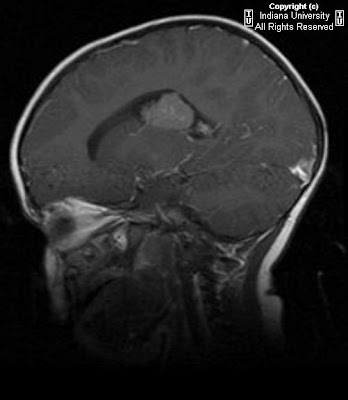
CT images demonstrate a mildly hyper attenuating intraventricular lobular mass arising within the lateral body of the right lateral ventricle. There is associated ventriculomegaly, indicative of at least mild hydrocephalus.
MRI images confirm the presence of a well-delineated, lobulated mass in the right lateral ventricle. Mass is iso intense to gray matter on T1WI and demonstrates heterogeneous enhancement following gadolinium-DTPA. Bright periventricular signal on axial FLAIR images likely represents mild transependymal interstitial edema due to hydrocephalus.

Differential diagnosis:
- Choroid plexus papilloma/carcinoma
- Intraventricular meningioma
- Papillary ependymoma


Diagnosis: Choroid plexus papilloma (WHO Grade I)


Key points

Choroid plexus tumors (CPTs) are intraventricular, papillary neoplasms derived from choroid plexus epithelium
One of the more common supratentorial brain tumors in children less than 2 years of age; it represents the most common intraventricular neoplasm in children
86% present by 5 years of age
WHO grade I

Location:
- 70% of CPTs occur in the atrium of the lateral ventricle (L>R)
- 20% occur in the 4th ventricle (most common site of origin in adults)

Imaging findings:
- NECT: iso- or hyper attenuating intraventricular lobular mass
25% have calcifications
Hydrocephalus is common due to ventricular obstruction and/or CSF overproduction
- CECT: Intense, homogeneous enhancement
- MRI:
T1WI: iso- to hypo intense well-delineated, lobulated intraventricular mass
T2WI: iso- to hyper intense +/- internal linear and branching flow voids
FLAIR: Bright periventricular signal due to transependymal interstitial edema
T1WI+C: Robust homogeneous enhancement; cysts and small foci of necrosis may be present

May spread via subarachnoid space
May become anaplastic over time; imaging alone cannot reliably distinguish between choroid plexus papillomas and carcinomas
Treatment is total surgical resection (5 year survival close to 100%)

giovedì 4 febbraio 2010

Septo-optic dysplasia


Findings

Absence of the septum pellucidum. Hypoplastic pituitary. Nonvisualization of the pituitary stalk. Hypoplasia of the optic nerves and optic chiasm.

Differential diagnosis:
- Septo-optic dysplasia
- Kallman syndrome
- Holoprosencephaly


Diagnosis: Septo-optic dysplasia


Key points

Also known as de Morsier's syndrome
Manifested by hypoplasia (underdevelopment) of the optic nerve, hypopituitarism and absence of the septum pellucidum
In a severe case, this results in pituitary hormone deficiencies, blindness, and mental retardation. However, there are milder degrees of each of the three problems, and some people only have one or two of the three.
Majority have other brain abnormalities as well, schizencephaly is most common
Ocular anomalies (coloboma, anophthalmia, microphthalmia)

Imaging findings:
- Absent septum pellucidum
- Flat roof of frontal horn, inferior aspect of frontal horn point down
- Small optic chiasm
- Thin pituitary stalk
- Posterior pituitary ectopia
- Callosal-forniceal continuation or fused midline fornices
- Thin corpus callosum
- Heterotopias, schizencephaly

mercoledì 3 febbraio 2010

Hemorrhagic meningoencephalitis


Findings

Figure 1: Coronal US image of the brain shows areas of cystic necrosis and hemorrhagic abscess.
Figure 2: Coronal US image of the brain shows ventriculomegaly.
Figure 3: Coronal US brain image shows periventricular cystic changes, parenchymal hemorrhage and abscess.
Figure 4: Coronal US brain image shows periventricular cystic change and a calcified hemorrhagic abscess.
Figure 5: Axial nonenhanced CT scan image of the brain shows multiple calcified hemorrhagic abscesses.
Figure 6: Axial CT scan image of the brain shows diffuse white matter destruction. The cortex is spared initially in the disease process.


Diagnosis: Bacillus cereus hemorrhagic meningoencephalitis


Group B beta-hemolytic streptococci and E coli are the most common causes of meningoencephalitis in the neonatal period. Serratia marcescens, or Citrobacter, may be considered as a causal micro-organism in cases of hemorrhagic meningoencephalitis. It is important to note that Bacillus cereus, though rare, can also cause hemorrhagic meningoencephalitis which is most often fatal. B cereus is a gram-positive, aerobic, spore-forming rod which is ubiquitous in the environment. The presence of this organism in blood or CSF is often regarded as a contaminant of specimens received by the microbiology laboratory and is therefore often disregarded by clinicians and microbiologists.

Acquisition of an infection by Bacillus cereus is thought to be nosocomial and is linked to the use of central or peripheral catheters, contaminated dressings, hospital linens, and ventilator equipment. Premature infants in ICU units are most vulnerable to this infection. Vasculitis, vasospasm, hydrocephalus, and diminished cerebrovascular autoregulation phenomena which are often seen in premature infants, are some of the risk factors for the infection.

Bacillus cereus is known to swarm out of the veins and cause extensive tissue damage and liquefactive necrosis by producing toxins, which include a necrotising enterotoxin, phospholipases, proteases, and haemolysins. In the brain, the destruction initially involves the subcortical and periventricular white matter extensively, sparing the cortex. The cortex can become involved in later stages and when this occurs, it preferentially involves the occipital lobes. Involvement of the basal ganglia is rare and can occur as a part of extensive brain destruction.

Brain sonograms obtained in the first 2-3 days can be normal but days later show asymmetrical white matter hyperechogenicity due to widespread destruction. This is later followed by development of multiple cysts, often with rims of hyperechogenicity around them. Multiple areas of parenchymal or intraventricular hemorrhage may also be demonstrated on ultrasound. Sometimes, brain sonography shows intraventricular septa and thickened ependyma due to ventriculitis. CT features include diffuse hypodensity of the white matter with multiple areas of hemorrhage and abscesses. The abscesses may undergo cavitatory necrosis and calcification. On MRI, multiple, confluent white matter hyperintensities are seen on T1W images and hypointensities on T2W images, both consistent with hemorrhagic destruction. DWI show restricted diffusion in the brain parenchyma due to cytotoxic edema and may also show lesions that were not detected on conventional MR images. The rate at which the findings develop sequentially in cases of suspected Bacillus cereus meningoencephalitis can be rapid and hence it’s worthwhile to consider serial sonography.


Differential diagnosis

PVL: PVL is an ongoing process which in many cases starts with symmetrical flaring in the periventricular white matter within the first days of life sometimes evolving into cystic destruction of the white matter. The condition is bilateral and almost always symmetrical, preferentially damaging posterior frontal and parietal regions. This can be confused with B cereus meningoencephalitis in the premature neonate. B cereus meningoencephalitis shows a dramatic change on serial sonograms performed whereas in PVL, the rate of evolution of the cystic changes is much slower. The presence of multiple abscesses in meningoencephalitis and the detection of the causative organism in the CSF may help differentiate the two conditions.

Asphyxia: shows transition from abnormal hyperechogenicity in major arterial areas to slow (in the course of a few weeks) destruction. This is in contrast to very rapid destruction of an initially normal brain in cases of bacterial encephalitis.

Herpes simplex viral encephalitis: The cortex and deep gray nuclei may also be affected, because of infarction caused by vasculitis and obstruction of small vessels, as well as neuronal apoptosis.

Deep cerebral vein thrombosis: Deep cerebral vein thrombosis often causes destruction of the white matter, the deep gray matter, basal ganglia, and thalamus. Moreover, thrombus is often visualized on US or CT involving the sagital sinus or the deep cerebral veins. The areas of destruction in B cereus are not compatible with arterial or venous infarction and do not correspond to arterial/ venous territories that would be involved in arterial or venous infarction.