lunedì 30 luglio 2007

Idiopathic dural dysplasia


Findings

Figure 1 and Figure 2: Axial and sagittal CT images demonstrate dural ectasia with a capacious thecal sac to the level of the sacrum.
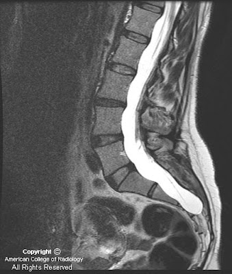
Figure 3 and Figure 4: Sagittal T1 postcontrast and T2-weighted MR images reveal a dilated terminal thecal sac without a tethered cord. Benign subtle scalloping of the posterior margin of the lumbar and sacral vertebral bodies (Figure 4) is best visualized on the T2-weighted images.
Figure 5 and Figure 6: Axial T1 postgadolinium and axial T2-weighted MR images reveal a dilated thecal sac with root sleeve prominence.

Differential diagnosis:
- Idiopathic dural dysplasia
- Neurofibromatosis I
- Marfan syndrome
- Ehlers-Danlos syndrome
- Homocystinuria
- Achondroplasia
- Hurler syndrome (MPS IH)
- Syringomyelia
- Ankylosing spondylitis


Diagnosis: Idiopathic dural dysplasia


This case is an example of an expanded dural sac with posterior vertebral scalloping of the lumbar spine and sacrum. Findings include a capacious thecal sac with smooth scalloping of the posterior aspect of the involved vertebral bodies. Dural dysplasia most often occurs in the lumbar spine but can involve the cervical and thoracic spinal canal as well. The case described herein is mild as there is no resulting kyphoscoliosis or erosion of the pedicles which occurs in more severe cases. Making the diagnosis of dural dysplasia requires that one excludes other causes of the expansion of the canal such as syrinx, tumor or meningeal cyst. The differential diagnosis and etiology of dural dysplasia is extensive and should be distinguished from meningeal cysts. The patient in this case had no known cause for the dural dysplasia and only complained of back pain.


Meningeal cysts
- Type I meningeal cysts: are extradural cysts that do not contain nerve root fibers. Type IA cysts are extradural arachnoid cysts. Occult sacral meningoceles (OIM) are considered type IB meningeal cysts which are also extradural and do not contain nerve root fibers. OIMs present with smooth remodeling and enlargement of the sacral canal with an extradural arachnoid sacral cyst adjacent to the thecal sac.
- Type II meningeal cysts are extradural cysts that contain nerve root fibers. These include Tarlov cysts and spinal nerve root diverticula. Tarlov cysts are cystic dilatation of the sacral root pouches with associated bone erosion which may or may not be symptomatic.
- Type III meningeal cysts are true intradural arachnoid cysts.

venerdì 27 luglio 2007

Arachnoid cyst with hemorrhage


Findings

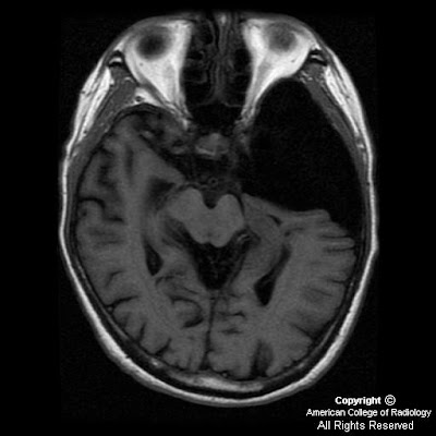
Figure 1: CT of the brain without contrast demonstrates a left extra-axial mass occupying the left frontotemporal region, which is relatively isointense to the white matter, measuring approximately 20 Hounsfield units.
Figure 2 : CT of the brain with bone windows demonstrates slight deformity of the calvarium with thinning related to a long-standing process.
Figure 3, 4, 5, 6, 7 and 8: Axial T1, T2, FLAIR and SPGR sequences demonstrate increased signal intensity compatible with subacute hemorrhage into left middle cranial fossa arachnoid cyst with left-to-right midline shift. Note that this is an atypical arachnoid cyst as it does not follow CSF on all sequences.
Figure 9: DWI image from original study obtained several months prior shows characteristic low signal consistent with uncomplicated arachnoid cyst.


Diagnosis: Arachnoid cyst with hemorrhage


Arachnoid cysts are cerebrospinal fluid (CSF) collections contained within a wall of normal arachnoid cells. These structures represent the most common intracranial congenital cystic lesions. They arise during development when the embryonic meninges fail to merge with resultant splitting of the arachnoid membrane. Importantly, they do not openly communicate with the ventricular system or subarachnoid space and typically show delayed opacification upon intrathecal contrast administration.

These lesions account for approximately 1% of all intracranial masses. There is a predilection for males (3:1) and they may be seen in any age group with 75% occurring in the pediatric population. Most cases are incidental findings in adult patients with brain imaging performed for unrelated symptoms. They usually do not enlarge over time however, can expand when CSF pulsations become entrapped in the arachnoid cyst.

When symptoms are present, they are related to the location and size of the lesion. Small cysts are typically asymptomatic whereas large masses present with various clinical features. The most common symptoms and signs include headache, seizures, developmental delay, hydrocephalus, and increased intracranial pressure. Focal neurological signs secondary to direct compression occurring less frequently.

These lesions demonstrate characteristic features on imaging studies as cystic cisternal masses with thin walls containing CSF density (CT) or intensity (MR) fluid. CT findings include a CSF attenuation mass (0 to 20 Hounsfield units) with sulcal effacement, displacement of surrounding structures, and remodeling or erosion of adjacent bone. There is no enhancement of the cystic contents or wall and calcification is rare. Occasionally, hemorrhagic products or proteinaceous fluid may result in higher attenuation and in these cases, MR is often the diagnostic modality of choice. On MR imaging, the extra-axial mass demonstrates signal intensity identical to CSF on all pulse sequences. Thus, it has low signal intensity on T1WI and high signal intensity on T2WI. Additionally, the FLAIR sequence shows a low signal (fluid-attenuated) lesion and diffusion-weighted imaging (DWI) also reveals a low intensity mass demonstrating absence of restricted diffusion.

Most cases do not require treatment and surgery is reserved for cases where symptoms correlate with anatomic location. Treatment options include conventional shunt placement for drainage into the peritoneal cavity or alternatively, cyst fenestration into normal CSF pathways (decompression) through an endoscopic approach or open craniotomy. Patients should be followed with serial scans for progressive cyst enlargement. As this case demonstrates, intracystic hemorrhage is a potential complication. Additional sequelae include secondary infection of the cyst or development of a subdural hematoma/hygroma.

giovedì 26 luglio 2007

Neurosarcoidosis


Findings

There is demonstrate diffuse, nodular meningeal thickening and enhancement along the convexities, interhemispheric fissures, and skull base. There is associated underlying parenchymal edema within the frontal lobes.

Differential diagnosis
- Granulomatous disease (namely, sarcoid)
- Meningitis
- Metastases
- Meningioma
- Histiocytosis


Diagnosis: Neurosarcoidosis (dural and parenchymal involvement)


Key points

Multisystem inflammatory disease characterized by noncaseating epithelioid-cell granulomas; etiology unknown
CNS involved in 5% clinically (27% autopsy); 10-20 per 100k in North America

Involves
- Dura, leptomeninges, subarachnoid space
- Brain parenchyma to include hypothalamus>brain stem>cerebral hemispheres>cerebellar hemispheres

Solitary or multifocal CNS mass(es)
Approximately 50% have periventricular T2 hyperintense lesions
Perivascular infiltrative involvement Virchow Robin spaces
May induce a small vessel vasculitis

Clinical Presentation:
- Most common symptom - CN deficit(s); most often CN VII; up to 50% asymptomatic
- Age of onset – 3rd-4th decade; 3-5% children; M:F 2:1; African American: Caucasian 10:1
- Pulmonary involvement in >90% of patients (abnormal CXR in association with CNS involvement strong evidence)

Course/Treatment:
- 2/3 have self limited monophasic illness; remainder have chronic remitting-relapsing course
- No known cure


Radiology

CT:
- May show basilar leptomeningeal enhancement
- Osteolytic skull lesions

MR:
- T1 + Contrast
Wide spectrum of enhancement
1/3 have multiple parenchymal lesions
>1/3 have leptomeningeal involvement, nodular and/or diffuse
10% solitary intra-axial mass
5-10% hypothalamus, infundibular thickening
- FLAIR:
Approximately 50% with periventricular T2 hyperintense lesions
Hyperintense vasogenic edema secondary to perivascular infiltrates or small vessel vasculitis
- T2:
Lacune (brainstem, BG)
Hypointense material within subarachnoid space
Hypointense dural lesion(s)
Hydrocephalus

giovedì 19 luglio 2007

Juvenile angiofibroma


Findings

CT shows a soft tissue mass extending from pterygopalatine fissure into sphenoid sinus with some erosion and expansion of adjacent bony structures. Angiography shows a major hyper vascular tumor supplied by sphenopalatine branch of right internal maxillary artery, without ascending pharyngeal artery supply. Minimal tumor blood supply from sphenopalatine branch of left internal maxillary artery. Super selective embolization of left and right sphenopalatine arteries was performed using Echelon-14 micro catheter and RVA particles (150-250 microns).


Diagnosis: Juvenile angiofibroma


Key points

Epidemiology:
- 1 of every 5,000-6,000 otolaryngological admissions.
- Approximately 0.5% of all head and neck neoplasms.
- Occurs exclusively in adolescent males.

Pathophysiology:
- Highly vascular tumors, locally invasive, non-encapsulated tumors.
- Usually arise at posterior attachment of middle turbinate, near the sphenopalatine foramen.
- Superior growth occurs towards sphenoid sinus, with erosion possible. Invasion into cavernous sinus may also occur.

Clinical:
- Symptoms: epistaxis (45-60%), nasal obstruction (80-90%), headache (25%), facial swelling (10-18%).

Treatment:
- Definitive therapy is usually surgical, with pre-operative embolization to control intraoperative bleeding.
- Hormonal therapy (with testosterone blockers) and radiotherapy have been tried with mixed results.


Radiologic overview

Usually suspected via findings on CT, but angiography used for definitive diagnosis and possible embolization prior to definitive surgery.
Most suggestive finding is a homogenous, nasopharyngeal soft tissue mass causing expansion of the nasal cavity, sometimes with septal deviation, and extending into the pterygopalatine fossa and sphenoid sinus.
Often see anterior bowing of the posterior wall of the ipsilateral maxillary sinus, but rarely with breakthrough into the antrum. Conversely, the tumor often extends superiorly with erosion into the sphenoid sinus, and possibly with extension into the cavernous sinus.
Angiography: 94% of the time, primary feeder system comes from branches of the external carotid system (usually from maxillary artery, but may also include ascending pharyngeal or vidian arteries).

mercoledì 18 luglio 2007

Sjogren syndrome involving the parotid glands


Findings

Figure 1 and Figure 2: There are diffusely enlarged, heterogeneous parotid glands with invasion of the carotid space and punctate calcifications in the left parotid gland. Discrete areas of low attenuation likely represent areas of parenchymal destruction or contained saliva.


Diagnosis: Sjogren syndrome involving the parotid glands


Sjogren’s syndrome is a chronic, systemic exocrinopathy secondary to lymphocytic infiltration of the salivary and lacrimal glands. Sjogren’s is the second most common autoimmune disorder after rheumatoid arthritis. Sjogren’s affects patients between 50-70 years of age with a 90% to 95% female predominance. Sjogren’s is primarily characterized by dry eyes (keratoconjunctivitis sicca) and dry mouth (xerostomia) although patients may develop symptoms referable to multiple organ systems similar to systemic lupus erythematosus. Certain systemic manifestations are unique to Sjogren’s syndrome such as interstitial nephritis, hyperglobulinemic purpura, and an increased risk of lymphoma. On the opposite end are patients complaining of dry eyes and mouth with vague symptoms of fatigue, myalgia, and cognitive dysfunction which may be difficult to distinguish from fibromylagia or depression.

The diagnosis of Sjogren’s syndrome involves both subjective and objective criteria. Patient’s complain of inadequate tear production and decreased saliva production. Ocular signs of corneal damage are present on physical exam (Schirmer’s or Rose Bengal) with a slit lamp exam. Laboratory diagnosis of Sjogren’s syndrome involves detection of antibodies to the Sjogren A or B antigen, SS-A (Ro) and SS-B (La). Labial biopsy and additional test indicating impaired salivary gland function may also be performed.

There are primary and secondary forms of the Sjogren’s syndrome. Primary Sjogren’s syndrome generally consists of dry eyes and mouth due to salivary and lacrimal gland destruction via autoimmune activation of lymphocytes. Secondary Sjogren’s syndrome, usually due to rheumatoid arthritis, causes symptoms related to exocrinopathy and collagen vascular disease. These patients have high titers of the SS-A antibody. Other secondary causes of Sjogren’s syndrome include systemic lupus erythematosus or scleroderma.

The imaging appearance of the parotid glands in Sjogren’s syndrome is varied and depends on the stage of the disease. The parotid glands may appear normal in the early stages. Multiple small cysts in both parotids can be seen in the intermediate form of the disease. Late stage Sjogren’s syndrome of the parotids involves large cystic and solid masses. The cystic components represent areas of destroyed gland or collections of saliva while the solid masses represent lymphoid aggregates which actively destroy the gland. Contrast enhanced CT typically demonstrates bilateral parotid enlargement, heterogeneous enhancement of solid and mixed cystic lesions and punctate calcifications. MR sialography is the best imaging test if Sjogren’s syndrome is suspected because of its ability to accurately stage the severity of the disease.

giovedì 12 luglio 2007

Corpus callosum lipoma, Agenesis of the corpus callosum, and Chiari I malformation


Findings

Nonenhanced axial CT demonstrates a well-defined, very low attenuation (fat) mass in the region of the roof of the third ventricle/genu of the corpus callosum. More inferiorly (Figure 2 and Figure 3), extension of the mass through the foramina of Monroe is noted (Figure 2). There are bilateral linear calcifications, which are classic for callosal lipoma (Figure 1, Figure 2, and Figure 3). In addition, there is parallel alignment of the lateral ventricles, suggestive of dysgenesis of the corpus callosum. There is also prominence of the occipital horns of the lateral ventricles (Figure 2), termed colpocephaly.
Axial T1 (Figure 4) weighted MR image through the center of the mass demonstrates a homogeneous very high signal mass in the region of the genu of the corpus callosum. Parallel orientation of the lateral ventricles is again seen. Incidentally noted is a large scalp lipoma.
Axial FLAIR (Figure 5) MR image in the same location demonstrates the callosal mass and the the scalp lipoma.
Sagittal T1 (Figure 6) weighted MR image in the midline demonstrates a fat intensity mass with absence of the corpus callosum. In addition, the cerebellar tonsils protrude approximately 5 mm below the level of the foramen magnum (blue arrow), consistent with a Chiari I malformation. Another scalp lipoma is also noted.


Diagnosis: Corpus callosum lipoma, Agenesis of the corpus callosum, and Chiari I malformation


CNS lipoma is an uncommon congenital lesion and constitutes less than 1% of brain tumors. Its most frequent location is the genu of the corpus callosum, tuber cinereum, and quadrigeminal plate cistern. The optic chiasm, interpeduncular cistern, sylvian fissure, cerebellopontine angle, and cerebellomedullary cisterns are less common locations.

CNS lipoma has the classic appearance of a discrete fatty mass. It often (especially when located within the corpus callosum) has a calcified rim, which can form a “bracket sign” on frontal radiographs, and this appearance is pathognomonic. It is very low attenuation on CT, homogeneously high in signal on T1- and T2-weighted MR images, and demonstrates no enhancement.

Approximately half of patients are asymptomatic at the time of discovery. Agenesis of the corpus callosum can be associated with colpocephaly, the disproportionate dilation of the occipital horns of the lateral ventricles compared to the frontal horns. This is felt by most to be due to decreased brain volume within the posterior fossa. However, colpocephaly and hydrocephalus can coexist, and the frontal and temporal horns should be carefully evaluated in the context of the clinical presentation.

There is a known association between callosal agenesis and other midline defects, including Chiari malformations, Dandy-Walker cyst, encephalocele, and others. A careful search should be undertaken for these congenital anomalies in any patient with callosal agenesis. In this patient, multiple scalp lipomas were also noted, raising the possibility of a lipomatosis syndrome.

Bilateral optic nerve glioma


Additional clinical history: Patient also has café au lait spots, and his mother has a history of Neurofibromatosis Type 1.


Findings

CT Head: There is enlargement of the bilateral optic nerves and optic chiasm.
MRI Brain: There is fusiform enlargement of the optic nerves bilaterally, with extension to the optic chiasm. The optic nerves demonstrate isointense signal to grey matter on T1 (and, not shown, also on T2 and FLAIR images), with contrast enhancement.

Differential diagnosis:
- Optic nerve sheath meningioma
- Optic neuritis
- Pseudotumor (Idiopathic orbital inflammatory disease)
- Adult malignant optic glioma
- Bilateral optic nerve glioma associated with NF-1


Diagnosis: Presumed bilateral optic nerve glioma in NF-1 patient


Key points

General:
- Most common cause of optic nerve enlargement
- Much more common than optic nerve sheath meningioma (up to 4 times more common)
- 3% of all orbital tumors
- Treatment may include radiation, chemotherapy, or surgery if needed
- Strong association with NF-1
Up to 50% of cases of optic nerve glioma involve patients with NF-1
Conversely, 15% of patients with optic nerve glioma have NF-1
May be bilateral in NF-1

Clinical Presentation
- Patients present with progressive vision loss
- May have proptosis
- Age at presentation is median of 5 years old
- Approximately 80% occur by 10 years of age
- Rarely progress after 6 years old, with cases of spontaneous regression

Pathology
- Grade I astrocytoma of the optic nerve
- Same histologic appearance as juvenile pilocytic astrocytoma


Radiology

Fusiform or sausage like enlargement of the optic nerve
Resulting in kinking of the optic nerve
May extend into proximal optic pathway, including optic chiasm, optic tracts, lateral geniculate body, and optic radiations
Demonstrates variable enhancement
Causes enlargement of optic canal

CT
- Isodense optic nerve enlargement
- Rare to have calcification (different from meningioma)

MR
- T1 iso/hypointense
- T2 variable signal
- Variable enhancement

mercoledì 11 luglio 2007

Graves ophthalmopathy


Findings

Figure 1: Axial CT-There is symmetrical proptosis on both sides. There is marked homogeneous enlargement of the muscle bellies of the extraocular muscles.
Figure 2: Coronal CT- Demonstrates enlargement of multiple muscles within the orbits bilaterally, with relative sparing of the lateral recti muscles.
Figure 3: Sagittal CT- There is crowding at the orbital apex with straightening of the optic nerve.
Figure 4: Axial postcontrast MRI – Homogeneous enhancement of the enlarged muscle bellies. There is marked bending of the lamina papyracea secondary to marked enlargement of medial recti muscles on both sides.


Diagnosis: Thyroid associated orbitopathy (Graves ophthalmopathy)


Thyroid associated orbitopathy (TAO), frequently termed Graves ophthalmopathy, is an autoimmune orbital inflammatory condition that is strongly associated with dysthyroidism. The lymphocyte-mediated inflammatory process affects the extraocular muscles, periorbital fat and connective tissues.

The eye findings associated with Graves disease can occur before, during, or long after the thyroid disease has been discovered or treated. While the orbitopathy is most commonly associated with hyperthyroid states, it can be seen in euthyroid and even hypothyroid patients.

It is associated with:
1) Graves hyperthyroidism (80%);
2) Hashimoto's thyroiditis (10% to 15%); or
3) unclassified thyroid immunologic abnormality (5%).

Thyroid associated orbitopathy usually affects young and middle-aged adults, females being affected 3 to 6 times more commonly than males. It may result in eyelid retraction, proptosis, chemosis, periorbital edema, and altered ocular motility with vision-threatening exposure keratopathy, troublesome diplopia, and compressive optic neuropathy occurring in untreated cases. TAO usually has a self-limited course, but significant chronic orbitopathy may occur in 10% to 15% of cases. Stable TAO can occasionally reactivate, but this is uncommon.

Orbital involvement is bilateral in 90% cases, although it may be asymmetric and symptoms may be unilateral. There is bilateral enlargement of extraocular muscles with increased orbital fat resulting in exophthalmos. The inferior and medial recti muscles are first to be involved. The lateral rectus muscle is the last to be involved and rarely shows isolated involvement. Muscle enlargement characteristically involves the belly, sparing the tendinous attachment to the globe.

Lacrimal gland enlargement may be seen. CT and MRI may also show “stretching” and apical crowding of the optic nerve with an enlarged superior ophthalmic vein.

TAO usually has a self limited course with favorable outcome. In approximately 10% of cases further therapy is required, such as systemic glucocorticoids or orbital radiotherapy. Surgical orbital decompression is reserved for those patients in which vision is threatened, where there is the presence of severe cosmetic deformity, or failure of medical management.

giovedì 5 luglio 2007

Joubert syndrome


Findings

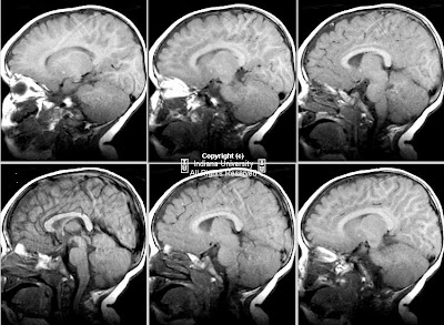
There is elevation of the fourth ventricle and hypoplasia of the superior portion of the vermis. There is thickening of the superior cerebellar peduncles bilaterally. These form a characteristic "molar tooth" sign as seen on axial images.


Diagnosis: Joubert syndrome


Key points

The syndrome was first described by Joubert and colleagues as a familial agenesis of the cerebellar vermis and appears to be inherited as an autosomal recessive trait. Both sexes are affected and the onset is in early infancy. Its incidence is unknown. Most patients die in infancy or early childhood. The predominant abnormality in Joubert's syndrome is aplasia or hypoplasia of the vermis, particularly the superior portion. In addition, these patients have heterotopic and dysplastic cerebellar tissue, abnormal development of the inferior olivary nuclei, and incomplete formation of the pyramidal decussation.


Clinical diagnosis

The most common features of Joubert's syndrome in infants include hyperpnea, jerky eye movements, and ataxia. The facial appearance in patients with Joubert syndrome may be near-normal, or may include high, rounded eyebrows, broadening of the nasal bridge, and mild epicanthus. The nares may be anteverted, and the mouth triangularly shaped.

Some patients with Joubert syndrome are severely affected, dying in infancy. The vast majority (> 90%) of surviving patients with Joubert syndrome are below chronological age in cognitive and motor development, although in a few patients, cognition may appear near-normal. CNS anomalies reported with Joubert's syndrome include callosal dysgenesis, congenital retinal dystrophy, and oculomotor abnormalities. Non-CNS disorders include neonatal breathing abnormalities, polydactyly, and cystic kidney disease.


Radiologic overview

Sagittal T1W images demonstrate a diminutive vermis. Axial images in particular show an enlarged fourth ventricle that is "bat-wing shaped" in configuration. The superior cerebellar peduncles are vertically oriented and elongated in the anteroposterior direction. Because of the dysgenesis of the vermis, the hallmark of Joubert's syndrome is separation or disconnection of the cerebellar hemispheres, which are apposed but not fused in the midline. The midbrain is small in its anteroposterior diameter, probably because of the absence of the decussation of the superior cerebellar peduncles. The characteristic appearance of the midbrain, with the enlarged superior cerebellar peduncles and the absence of their decussation has been called the "molar tooth sign". Associated supratentorial anomalies are uncommon, but cerebral cortical dysplasia and gray matter heterotopia have been reported.


Management and treatment

The prognosis for infants with Joubert's syndrome depends on whether or not the cerebellar vermis is entirely absent or partially developed. Some children have a mild form of the disorder, with minimal motor disability and good mental development, while others may have severe motor disability and moderate mental retardation. Treatment for Joubert syndrome is symptomatic and supportive. Infant stimulation and physical, occupational, and speech therapy may benefit some children. Infants with abnormal breathing patterns should be monitored.


mercoledì 4 luglio 2007

Ependymal cyst


Findings

Figure 1: Axial T1 weighted image shows a large well defined homogenous mass in the right lateral ventricle which is isointense with CSF and arises from the atrium and measures 8.8 cm by 4.2 cm by 6.0 cm. There is dilatation of the right occipital horn and midline shift to the contralateral side indicating mass effect.
Figure 2: Axial T2 weighted image demonstrates that the mass is again isointense with CSF, indicative of cyst. The thin regular cyst wall is visualized. There is no edema in the adjacent brain parenchyma.
Figure 3: Diffusion-weighted image illustrates no evidence of restricted diffusion. The fluid within the cyst has the same diffusion characteristics as CSF.
Figure 4: Axial T1 weighted postgadolinium image shows no abnormal enhancement. Note the compressed vasculature adjacent to the cyst.
Figure 5: Coronal T1 weighted post gadolinium image depicts the large cyst in the right lateral ventricle.


Diagnosis: Ependymal cyst


Ependymal cysts are rare intracranial lesions that are found in the brain parenchyma, ventricles, and subarachnoid space. The cysts are most often juxtaventricular in location within the brain parenchyma involving the frontal or temporal lobes. They are less often found within the ventricles with the lateral ventricles being the most common. Subarachnoid ependymal cysts are extremely rare. The origin of the cysts is unknown, but they are thought to originate from invagination of neuroectoderm, and the cysts are lined with either cuboid or columnar epithelium.

Ependymal cysts are typically found in young males in their 30s and 40s. The cysts are usually asymptomatic and are incidental imaging findings, but when symptomatic, patients will present with headache, seizures, or dementia. On CT imaging, the thin cyst wall may not be visible, and the cystic fluid is of the same density as CSF. There is no enhancement following contrast administration. On MR imaging, the cystic fluid will be isointense to CSF on all pulse sequences. Sometimes the cyst may have a proteinacous content, due to mucinous secretion by the epithelial cells that line the cyst, which is represented by an increase in signal intensity best seen on FLAIR images. The cyst wall is often visible on MR, but its absence does not exclude the diagnosis. The cyst will not show restricted diffusion, and similar to CT, there will be no enhancement following contrast administration. Ependymal cysts of widely variable sizes have been reported, and large cysts may obstruct CSF flow and cause hydrocephalus.

Prognosis for ependymal cysts is excellent. Since most cysts are asymptomatic, patients can be followed conservatively. Cysts that are symptomatic require either surgical decompression or excision. Recurrence after surgical excision is rare.

The patient in this case is particularly interesting due to his extremely young age and symptomatic presentation. He was managed surgically with placement of a cystoperitoneal shunt catheter and has been seizure free since his hospital discharge.