


Findings
Axial CT shows elongated and thickened superior cerebellar peduncles. Note hte "bat-wing" appearance of the fourth ventricle.
Axial T2-weighted image (Figure 2) through again demonstrates characteristic "bat-wing" appearance of the fourth ventricle with enlarged superior cerebellar peduncles.
Axial T1-weighted inversion recovery image (Figure 3) at the level of the cerebellar peduncles again shows the enlarged superior cerebellar peduncles. The isthmus of the brainstem (the transitional zone between the pons and midbrain) is small and in combination with elongated thickened superior cerebellar peduncles produces the "molar tooth sign." Note the cerebellar hemispheres are in apposition without evidence of fusion.
Mid-sagittal T1-weighted MR (Figure 4) confirms absence of the cerebellar vermis.
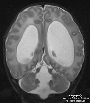
Coronal T2-weighted image (Figure 5) shows the thickened cerebellar peduncles (Figure 5) and absence of the normal vermis, without cerebellar hemispheric fusion.
Diagnosis: Joubert syndrome
Joubert syndrome is an autosomal-recessive disorder, characterized by clinical presentation of hypotonia, ataxia, and global developmental delay. A variety of other abnormalities have been described in affected children, primarily episodic hyperpnea, abnormal eye movements, and a characteristic facial appearance. The phenotype may vary even among siblings with Joubert syndrome.
From an imaging perspective, these patients have either complete or partial agenesis of vermis, which results in triangular-shaped mid fourth ventricle and a bat-wing appearance in its superior aspect. The cerebellar hemispheres oppose one another in the midline due to absence of vermis. The isthmus and the midbrain are small in AP diameter, likely secondary to absence of decussation of the superior cerebellar peduncles. This appearance of small brainstem with elongated and thickened superior cerebellar peduncles has been termed the molar tooth sign. It has been proposed that both the superior cerebellar peduncles and the corticospinal tracts remain uncrossed in these patients. The classic imaging findings are, however, not completely specific for Joubert syndrome and have been found recently in a number of very rare congenital syndromes.
Thankyou for posting this
RispondiEliminaDr. Arvind Nanda provides one of the best brain hemorrhage treatment in Delhi and he offers interventional radiology treatments for the brain hemorrhagic stroke.