giovedì 29 novembre 2007
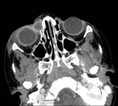
Dacryocystitis
Findings
Ring enhancing lesion in the region of the right lacrimal sac, contiguous with the nasolacrimal duct. Surrounding preseptal edema. Scattered relatively mild ethmoid air cell mucosal thickening and bilateral maxillary sinus mucosal thickening. Bones are unremarkable. A mucous retention cyst is seen in the nasopharynx.
Clinical differential diagnosis:
- Cellulitis
- Abscess
- Dacryocystitis
Diagnosis: Dacryocystitis
Stagnation of tears in a pathologically closed lacrimal drainage system can result in dacryocystitis.
Acquired dacryocystitis can be acute or chronic.
There is also a form of congenital dacryocystitis, thought to be related to lacrimal excretory system embryogenesis, specifically incomplete canalization of the nasolacrimal duct.
Acute dacryocystitis is manifested by the sudden onset of pain, erythema, and edema overlying the lacrimal sac region.
Complications include abscess formation and spread of infection into the adjacent orbit.
Etichette:
AuntMinnie,
Head - Neck,
Inflammatory
mercoledì 28 novembre 2007
Medulloblastoma
Findings
Large solid and cystic midline heterogeneously enhancing cerebellar mass effacing the fourth ventricle and causing lateral and third marked ventriculomegaly with transependymal edema. Two foci of leptomeningeal contrast enhancement in the posterior fossa, possibly metastases.
Differential Diagnosis:
- Pilocytic astrocytoma
- Medulloblastoma
- Ependymoma
- Brainstem glioma
Diagnosis: Medulloblastoma, undifferentiated type (WHO Grade IV)
Discussion
Medulloblastoma is somewhat more common in males than females with a ratio of approximately 1.5 to 1. This tumor accounts for approximately 7-8% of all intracranial tumors but over 30% of pediatric brain tumors. 75% of the cases occur in children, but the tumor can be seen at any age. The median age of presentation is 9 years. Presentation usually is secondary to hydrocephalus which develops because of mass effect from the tumor on the normal cerebrospinal fluid pathway. Truncal ataxia and trouble walking can be seen secondary to tumor invasion in to the cerebellum. Medulloblastomas can be seen in syndromes such as Gorlin's and Turcot's syndromes.
Medulloblastoma is a primitive neuroectodermal tumor which means it is a small round blue cell tumor. These types of tumors typically are tightly packed which results in a higher density than surrounding soft tissues. This fact helps with the diagnosis as these tumors will appear dense on noncontrasted head CT. This tumor will typically intensely and homogenously enhance. Calcification can be seen but it is uncommon.
Medulloblastoma can become metastatic via CSF spread. This a poor prognostic factor. Preoperative MRI can help identify metastatic lesion so they can be removed at initial surgery if possible. Surgery is the first line therapy for medulloblastoma. Postoperative chemotherapy and radiation are employed as well. The radiation therapy is extensive with the entire spine and brain being irradiated. With the combination of these three therapies the patients can reach an 80% 5-year survival rate excluding the metastatic at time of diagnosis patients.
Etichette:
AuntMinnie,
Neoplasm,
Neuro,
Pediatric
mercoledì 21 novembre 2007
Hydranencephaly
Findings
Figure 1: Noncontrast CT of the head reveals complete absence of supratentorial brain tissue which is replaced by cerebrospinal fluid. The falx is present.
Figure 2: Noncontrast CT of the head reveals complete absence of supratentorial brain tissue with residual pons and cerebellum.
Figure 3: Axial T2 reveals complete absence of supratentorial brain tissue which is replaced by cerebrospinal fluid. The falx is present.
Figure 4: Axial T2 reveals complete absence of supratentorial brain tissue with residual pons and cerebellum.
Figure 5: Sagittal T1 reveals the presence of the tentorium cerebelli and structures of the midbrain along with previous findings.
Diagnosis: Hydranencephaly
Hydranencphaly is a congenital malformation of the brain in which there is near total to complete absence of the cerebral cortex and basal ganglia. There is usually preservation of the thalami, cerebral peduncles, pons, midbrain and cerebellum. Although the main etiology is unknown, it is thought to be secondary to an in-utero event. Possibilities include infection (CMV, HSV, Toxoplasmosis), vascular accident, specifically occlusion of the anterior circulation, or diffuse hypoxic-ischemic necrosis due to maternal exposure to carbon monoxide or butane gas.
Hydranencephaly can be difficult to distinguish from extreme hydrocephalus, alobar holoprosencephaly and porencephaly. The presence of residual cerebral cortex helps to distinguish hydranencephaly from the other diagnoses. The absence of the third ventricle and presence of the falx also aids in differentiation since the third ventricle is present in the other diagnoses and the falx is absent in holoprosencephaly.
In utero ultrasound is helpful in diagnosing hydranencephaly. This usually presents as a large cystic mass filling the entire cranial cavity with absence or discontinuity of the cerebral cortex. The appearance of the thalami and brainstem protruding inside a cystic cavity is also characteristic. CT and MRI findings are similar with the absence of most supratentorial structures and preservation of the brainstem and cerebellum. MRI is superior for the detection of cortical remnants.
Prognosis in these children is very poor with most deaths occurring within the first year. Although children with hydranencephaly may appear healthy, developmental delay occurs within a few weeks. Irritability, abnormal muscle tone and seizures are common manifestations.
Etichette:
ACR,
Malformations,
Neuro,
Pediatric
Dural sinus thrombosis, with involvement of the superior sagittal sinus, straight sinus, and vein of galen
Findings
CT: There is marked dilation of the superior sagittal sinus with low attenuating density, containing high density septations. The expansion and density extends into the straight sinus. There is a large peripherally calcified lesion at the right aspect of the dilated superior sagittal sinus near the torcular Herophili. There is also severe hydrocephalus, with involvement of the third and lateral ventricles.
MRI: There is a heterogeneous lesion expanding the superior sagittal sinus and torcula, demonstrate mostly hyperintense signal on T1 and T2, containing multiple septations, with suggestion of fluid/fluid level, without definite contrast enhancement. There is extension into an enlarged vein of Galen. There is a area of low signal on the right side corresponding to calcification seen on CT.
No central flow is demonstrated in the posterior portion of the superior sagittal sinus, corresponding to the lesion.
Angiography (images not shown): Showed mass-effect in the posterior parietal-occipital regions without filling by contrast. No evidence of tumoral blush, AV shunting, or aneurysm.
Follow-up CT: Showed significant decrease in mixed density material expending the superior sagittal sinus from the vertex to the torcula, consistent with interval resorption of clot.
Differential diagnosis for the neonatal studies:
- Dermoid
- Vascular tumor
- Congenital variant
- Giant dural sinus thrombosis
Diagnosis: Dural sinus thrombosis, with involvement of the superior sagittal sinus, straight sinus, and vein of galen
Diagnosis confirmed on the subsequent CT, which demonstrated partial resorption of thrombus.
Discussion
Dural sinus thrombosis represents formation of clot in the dural sinus
Clot spreads from dural sinus to cortical veins, resulting in hemorrhage and infarct
Will progress to infarction in approximately 50%
Clinical presentation:
- Presenting symptoms include headache, focal neurologic deficits, papilledema
- Numerous etiologies, such as hypercoagulable state, infection, inflammation
- Dehydration and infection is a common cause in children
- Idiopathic in 10 to 30% of cases
- No particular age or sex predilection
- Morbidity and mortality from progression to venous infarction
- Treatment include thrombolysis
Imaging:
- Early findings are subtle
- Classic sign is the "empty delta" seen on contrasted imaging
- Thrombus seen in dural sinus and cortical veins
- Sinus is occluded and expands
- Appearance depends on age of clot
- Vessels may recanalize
CT:
- "Empty delta" sign represents non enhancing thrombus in the dural sinus with surrounding enhancing dura in contrast enhanced CT
- "Cord sign" is hyperdense dural sinus clot seen in non contrasted CT
- May see associated hemorrhage or edema
MRI:
- Signal on T1 and T2 depends on age of thrombus
- On GRE, the thrombus may "bloom," depending on stage of hemorrhage
- On MRV, there is absence of flow
Angiography:
- Thrombosed sinus does not fill
martedì 20 novembre 2007
Shaken-baby syndrome
Findings
Bilateral orbital hyper densities (retinal hemorrhage), loss of grey-white matter differentiation and effacement of the basal cisterns and ventricles (hypoxic ischemic injury, cerebral edema, impending herniation), left convexity cortical contusions / SAH, interhemispheric SDH.
Differential diagnosis:
- Non-accidental trauma
- Accidental trauma
- Hemophilia
- Hypoprothrombinemia
- Glutaric aciduria
- Osteogenesis imperfecta
- Leukemia
Diagnosis: Shaken-baby syndrome (non-accidental trauma)
Key points
The constellation of retinal hemorrhage, SDH, and long-bone metaphyseal fractures were initially described by Caffey as "whiplash shaken injury." Non-accidental trauma is the most common cause of traumatic death in infancy.
Risk factors: Prematurity, < 12 mos, young parents, low socioeconomic status, twin, male, disability.
Perpetrators: father > boyfriend > babysitter > mother.
Common presentation: "unresponsive" – upper cervical cord stretching causing apnea causing ischemic brain injury (60% mortality if comatose at presentation).
Neurologic manifestations include:
- SDH (highly specific if of various ages/locations, convexities / posterior interhemispheric fissure common).
- Retinal hemorrhage (usually bilateral, always associated with SDH).
- Cortical contusions.
- SAH
- Hypoxic ischemic encephalopathy with loss of grey-white differentiation and overall decreased cerebral attenuation with sparing of posterior fossa; effacement of sulci/ventricles/cisterns.
- Skull fractures and EDH are uncommon.
Plain film is more sensitive that CT in detecting skull fractures.
Skeletal survey should always be obtained if NAT is suspected.
Differential Diagnosis for retinal hemorrhage w/ SDH:
- accidental trauma (consider concordance of history given vs. severity of injury)
- coagulopathies (blood dyscrasias, leukemia)
- resuscitative efforts (retinal hemorrhage only)
- osteogenesis imperfecta (skeletal manifestations predominate)
- inborn errors of metabolism (glutaric aciduria type I associated with SDH only)
Etichette:
AuntMinnie,
Neuro,
Pediatric,
Trauma
venerdì 16 novembre 2007
Intraosseous meningioma
Findings
Figure 1 and Figure 2: Bone windows from axial CT images reveal expansion and sclerosis of the left sphenoid bone.
Figure 3: Axial CT section on brain windows demonstrates displacement of the optic nerve medially with bony expansion and an associated soft tissue component.
Figure 6, Figure 7 and Figure 8: T2, Axial FLAIR and gradient echo demonstrate expansion and diffuse low signal in the left sphenoid.
Figure 9 and Figure 10: Axial and coronal T1-weighted contrast enhanced images demonstrate minimal left sphenoid enhancement (Figure 7) and a conspicuous avidly enhancing, dural-based left temporal mass consistent with a meningioma.
Differential diagnosis:
- Intraosseous meningiomas
- Paget disease
- Fibrous dysplasia
- Sclerotic metastases
- Inflammatory lesions of the skull base
Diagnosis: Intraosseous meningioma
Meningiomas without a dural component are considered ectopic and can occur in a variety of locations including the subcutaneous tissues, paranasal sinuses, orbit, neck, salivary glands, the perineural sheath of cranial nerves and the calvaria. Purely intraosseous meningiomas (without an associated dural component) can be mistaken for osseous pathology including fibrous dysplasia, Paget disease and sclerotic metastases. Inflammatory lesions of the skull base, such as aspergillosis, may also give a similar appearance and should be considered in the proper clinical setting (sinus disease, diabetes, immunocompromise).
Meningiomas arise from arachnoid meningothelial “cap” cells. The trapping of “cap” cells in the cranial sutures during birth and subsequent modeling of the infant skull is thought to produce intraosseous meningiomas. This explains their predilection for the cranial sutures with the bony orbit and frontoparietal skull most commonly involved. Other proposed etiologies of intraosseous meningiomas include antecedent trauma, resulting in dural trapping within fracture lines and the multipotent nature of mesenchymal cells which give rise to the meninges and a variety of tissue types (including bone) which have been identified within meningioma pathologic specimens.
CT findings of meningiomas include a hyperdense (75%) smooth mass abutting the dura. Hyperostosis is a common occurrence in meningiomas; this may be secondary to local calvarial invasion, reaction to the adjacent dural mass or a primary osseous lesion. Calcification is identified in 25% of meningiomas which may cause focal areas of low signal intensity on MR imaging. The T1 weighted MR imaging appearance of meningiomas is an isodense extraaxial mass with a dural base and gray matter buckling. The appearance of meningiomas on T2 weighted images is isointense to cortex with a rim of CSF or trapped CSF clefts. Occasionally meningiomas may appear high in signal on T2 weighted imaging, indicating syncytial or angioblastic histology. Visualized flow voids may also be present within larger meningiomas, implying their vascularity. More than 95% of meningiomas demonstrate avid, uniform enhancement on post-contrast images. Meningiomas may display varying degrees of associated parenchymal edema.
Like their dural counterpart, purely intraosseous meningiomas demonstrate a 2:1 female predominance (in the 36 reported cases) and are generally considered benign. Intraosseous meningiomas demonstrate diffuse sclerosis and bony expansion on CT images. The MR appearance of intraosseous meningiomas is diffuse low signal of the expanded bone, the presence of an associated enhancing dural component greatly aids in the diagnosis by excluding both primary bone pathology and metastases. In meningiomas with both dural and intraosseous components, it is impossible to determine the site of origin as the osseous involvement may be the result of secondary invasion. Intraosseous meningiomas are treated with surgical excision when symptomatic. Cranial reconstruction with bone grafting is often necessary. Any remaining intraosseous component, secondary to involvement of sutures should be followed with serial radiographic examinations with adjuvant radiation for recurrent symptoms or lesion progression.
Etichette:
ACR,
Head - Neck,
Neoplasm,
Neuro
mercoledì 14 novembre 2007
Rhombencephalosynapsis
Findings
Fusion of the cerebellar hemispheres with partial absence of the vermis
Diagnosis: Rhombencephalosynapsis
Rhombencephalosynapsis is a unique posterior fossa malformation. The diagnosis is a very uncommon one with ~30 cases in the literature. The patients can present at nearly any age because of the broad range of clinical findings: from normal cognition to severe mental retardation and cerebral palsy. Common associated anomalies include fusion of the dentate nuclei, superior cerebellar peduncles and thalami, absence of septum pellucidum, septo-optic dysplasia, anomalies of limbic system and secondary hydrocephalus.
Etichette:
AuntMinnie,
Malformations,
Neuro,
Pediatric
giovedì 8 novembre 2007
Dandy Walker malformation
Findings
There is a large fourth ventricle which appears to be in continuation with a large posterior fossa cyst which follows CSF signal.
Differential Diagnosis:
- Arachnoid cyst
- Dandy walker variant
- Dandy walker malformation
Diagnosis: Dandy Walker malformation
Key points
Classic triad makes up the Dandy-Walker malformation
- Vermian agenesis
- Communicating with an enlarged 4th ventricle
- Posterior fossa enlargement
Dandy-Walker malformation may be isolated or can occur with other malformations of the brain.
Associated CNS abnormalities include
- Agenesis of the corpus callosum
- Lipomas
- Aqueductal stenosis
- Microcephaly
- Encephalocele
The Dandy Walker Spectrum represents a broad spectrum of cystic posterior fossa malformations.
These include (from most severe to least severe):
- 4th ventriculocele: DWM in which large posterior fossa cyst erodes occipital bone causing posterior outpouching of the cyst (encephalocele) through the occipital bone
- "Classic" Dandy Walker Malformation: Cystic dilatation of the 4th ventricle which opens dorsally to a variable sized CSF cyst which causes an enlarged posterior fossa and superiorly rotated hypoplastic vermian remnant; hydrocephalus is common
- Dandy Walker Variant: 4th ventricle opens to a smaller posterior cyst with partial rotation of the vermis, variable vermian hypoplasia; normal sized posterior fossa
- Persistent Blake Pouch Cyst: Open 4th ventricle communicates with persistent cyst that fails to regress and obliterates basal cisterns; normal vermis
- Mega Cisterna Magna: Enlarged cisterna magna, 4th ventricle is closed; normal vermis
DWM commonly presents with macrocephaly and bulging fontanel, 80% are diagnosed by 1 year of age. Early death is common.
Etiology is from hindbrain development arrest. No 4th ventricle outlet foramina are formed.
Associated with other CNS/extracranial abnormalities two-thirds of the time, such as craniofacial, cardiac and urinary tract abnormalities, polydactyly, orthopedic, and respiratory abnormalities
Treatment includes CSF diversion of hydrocephalus with VP shunt
Etichette:
AuntMinnie,
Malformations,
Neuro,
Pediatric
lunedì 5 novembre 2007
Subacute combined degeneration
Findings
Figure 2 and Figure 4: Sagittal and axial T2-weighted images demonstrate symmetrical hyperintensity within the dorsal columns that is contiguous in length over several vertebral body lengths.
Figure 1 and Figure 3: Sagittal T1-weighted images pre and post-gadolinium demonstrate hypointense signal within the dorsal cervical spinal cord without enhancement.
Diagnosis: Subacute combined degeneration
MR findings in subacute combined degeneration include symmetrical increased signal intensity in T2-weighted sequences along the dorsal columns that is usually contiguous over multiple vertebral body lengths.
The differential diagnosis includes B12 deficiency with subacute combined degeneration, tabes dorsalis and vacuolar myelopathy in AIDS.
Vitamin B12 deficiency causes a wide range of neurological, hematological and gastrointestinal disorders. Subacute combined degeneration is one such neurological disorder that is characterized by symmetrical numbness, weakness and paresthesia. It most commonly results from pernicious anemia and is potentially reversible with B12 supplementation.
MR findings include symmetrical increased signal intensity on T2-weighted sequences along the dorsal columns that is usually contiguous over multiple vertebral body lengths. Lesions are iso or hypointense to the normal cord on precontrast T1-weighted sequences. There can be variable contrast enhancement. The differential diagnosis includes tabes dorsalis and vacuolar myelopathy in AIDS. These can be distinguished from other myelopathies on the bases of their symmetry and contiguous cord level involvement. They can be differentiated from each other on the basis of laboratory tests and clinical history.
Etichette:
ACR,
Neuro,
Spine,
Toxic-Metabolic
Iscriviti a:
Post (Atom)