mercoledì 30 settembre 2009

Cholesteatoma


Findings

Non contrasted temporal bone CT reveals a soft tissue mass of the right tympanic membrane and Prussak’s space with associated erosions of the scutum, epitympanum walls, lateral semicircular canal, tegmen tympani and middle ear ossicles.

Differential diagnosis:
- Chronic otomastoiditis
- Acquired cholesteatoma
- Middle ear cholesterol granuloma
- Glomus tympanicum paraganglioma


Diagnosis: Cholesteatoma


Key points

An acquired cholesteatoma is a collection of exfoliated squamous epithelium and debris.
Cholesteatomas often begin at the pars flaccida of the tympanic membrane and grow in Prussak's space of the temporal bone and produce inflammatory reaction.
Cholesteatomas typically occur in the setting of chronic dysfunction of the Eustachian tubes and recurrent otitis media.
Cholesteatomas can be as small as a couple of millimeters in size or can grow to fill the entire middle ear.
Erosions of the scutum and ossicles are commonly seen.
The presence of bony erosions or expansion are strong support of cholesteatoma over chronic otomastoiditis.
Acquired cholesteatomas occur in children and adults.
Most common presenting symptoms include ear discharge, hearing loss, ear pain and vertigo.
Without treatment cholesteatomas will progressively increase in size.
Complications include: Hearing loss, CN 7 palsy, venous sinus thrombosis, semicircular canal fistulas, and intracranial invasion.
Early surgical intervention usually results in complete eradication and preservation of hearing.

venerdì 25 settembre 2009

Mineralizing microangiopathy


Findings

Figure 1: Both basal ganglia and thalamus show homogeneous high signal.
Figure 2: There is low signal within the medial aspect of the lentiform nucleus (globus pallidus) and caudate heads bilaterally.
Figure 3: There is high density material within the basal ganglia and subcortical white matter.
Figure 4: There is high density bordering the subcortical white matter of both the frontal and parietal lobes.
Figure 5: There is high density bordering the subcortical white matter of the frontal lobes and caudate nucleus.
Figure 6: There is increased density within the subcortical white matter of the frontal and parietal lobes at the centrum semi ovale.


Diagnosis: Mineralizing microangiopathy


Mineralizing microangiopathy is a distinctive histopathologic process involving the microvasculature of the central nervous system. Pathologically there is hyalinization and fibrinoid necrosis of small arteries and calcium deposition. It is usually seen following combined radiation and chemotherapy for the treatment of CNS neoplasm in childhood, although it can be seen in patients receiving only radiation. There is no direct correlation between the extent of the radiological findings and patient’s symptoms. However age appears to be important as most cases have been reported in patients receiving treatment before 10 years of age.

Radiological manifestations include parenchymal calcification within the basal ganglia, specifically the putamen and thalamus. There might be involvement of adjacent white matter tracts, cerebral or cerebellar cortex but this is less likely. MRI evaluation may show high signal on T1 images and low signal on T2 images within the above mentioned areas corresponding to calcifications. Although there is usually low signal on both sequences due to a paucity of protons, high signal can be explained by a surface relaxation effect by the calcium salt particle precipitated in the brain. Differentiating treatment induced abnormalities from residual or recurrent tumor is often difficult and requires close clinical and radiological correlation.

Histologically, the calcifications seen in mineralizing microangiopathy are present within the walls of precapillary arterioles, capillaries, venules, and smaller arteries such as the lenticulostriate. Involvement of adjacent white matter tracts, cerebral cortex and cerebellar structures has also been described but is less common.

martedì 22 settembre 2009

Intramedullary lipoma


Findings

Figure 1: Intramedullary mass at the T9-T10 level with hyperintense signal on T1-weighted sequence.
Figure 2: Intramedullary mass at the T9-T10 level with hyperintense signal on the T2 -weighted sequence.
Figure 3: Fat suppression on the STIR image.
Figure 4 and Figure 5: Fat saturation on the T1 fat saturated post contrast images and no enhancement of the lesion. There also is splaying of the spinal cord at this level.


Diagnosis: Intramedullary lipoma (surgically proven)


99% of intramedullary lipomas are associated with spinal dysraphism. Most tumors occur in the dorsal cervical and thoracic cord.

Intramedullary lipomas are rare. These comprise 1-2 % of intramedullary tumors. Multiple noncontiguous intramedullary lipomas are even rarer. These affect both sexes equally. Most patients present with symptoms during the first 2 -3 decades of life. Most have a slow progression of neurological deterioration. Sensory disturbances, pain, ataxia and lower extremity weakness are the most common presenting complaints. Urinary and bowel incontinence present later. CT can show a fatty lesion and MRI is diagnostic. They have a short T1 relaxation time and are bright on T1 weighted images and also bright on T2 weighted images following signal intensity of subcutaneous fat. There is very little enhancement. Treatment is still controversial and some physicians advocate early decompression before onset of symptoms because of significant residual deficits after surgery in symptomatic patients. Complete resection is not possible in intramedullary lipomas.

lunedì 21 settembre 2009

Subependymal giant cell astrocytoma


Findings


Axial T2 weighted image demonstrates a well-marginated intraventricular mass near the foramen of Monro. The lesion is lobulated in appearance and is causing obstructive hydrocephalus. Associated vasogenic edema is noted extending into the left frontal lobe. Axial and Coronal T1 contrast-enhanced MR sequences demonstrate intense, heterogeneous enhancement of the mass.

Differential diagnosis:
- Astrocytoma
- Subependymoma
- Germinoma
- Choroid plexus tumor
- Hamartoma


Diagnosis: Subependymal giant cell astrocytoma


Key points

Subependymal giant cell astrocytomas are seen as an enhancing intraventricular mass in a patient with tuberous sclerosis complex. Subependymal giant cell astrocytomas occur in about 10 percent of patients with TS.
Almost always located near the foramen of Monro.
Well marginated, often lobulated
Heterogeneous, strong enhancement
Growth favors the diagnosis (vs static tuber).
Brain MR with contrast should be recommended every 1-2 years for followup.
FLAIR sequence is useful for detecting subtle CNS findings of tuberous sclerosis.
SGCTs are pathologically indistinguishable from subependymal nodules, and radiologic criteria for distinguishing the lesions are unreliable. Clinical criteria are therefore more useful in decision-making regarding operative therapy.

venerdì 18 settembre 2009

Hydranencephaly


Findings

There is near complete absence of supratentorial brain parenchyma, which is replaced with CSF, with an intact falx. The brain stem and cerebellum are present and grossly normal in appearance.

Differential diagnosis:
- Hydranencephaly
- Severe hydrocephalus


Diagnosis: Hydranencephaly


Key points

Hydranencephaly - In utero destruction of cerebral parenchyma with intact falx and preservation of posterior fossa structures.
Cerebrum replaced with CSF.
Caused by in utero occlusion of bilateral supraclinoid internal carotid arteries. Etiology unclear (hereditary thrombophilic states, infection, maternal irradiation/toxin exposure, twin-twin transfusion, intrauterine anoxia).
Rarely unilateral
Occurs approximately <1:10000 births, greatest incidence in teenage mothers.
Clinically, present with macrocephaly, developmental delay, irritability, hyperreflexia, seizures.
Often seen with prenatal ultrasound (anechoic cranial vault).
On CT and MR (best characterized with MR) – CSF attenuation/signal intensity replacing the supratentorial brain parenchyma with sparing of thalami, brain stem, cerebellum, and choroid plexus.
Falx is intact (distinguishes from holoprosencephaly).
No thin rim of cerebral parenchyma (distinguishes from severe hydrocephalus).
Prognosis is poor – usually death in infancy.
Treatment is supportive care, shunting to decrease head growth.

giovedì 17 settembre 2009

Castleman disease


Findings

A left parotid lesion appears hyperintense on T1 and T2 (Figure 1 and Figure 3) as compared to muscle and demonstrates moderate to intense enhancement (Figure 4). The adjacent mandible is normal. Bilateral cervical lymphadenopathy (Figure 6 and Figure 8) is seen on T1 and T2 images, deep to the sternocleidomastoid muscle. Contrast enhancement within these lymph nodes is present (Figure 9).


Diagnosis: Castleman disease


Castleman disease (angiofollicular lymph node hyperplasia) is considered a benign lymphoproliferative disorder of unknown etiology and was originally described at the Massachusetts General Hospital by Dr. Benjamin Castleman in 1957. Distinction between the two subtypes – hyaline vascular or plasma cell – can be determined histologically. The hyaline vascular subtype is unicentric, more common, and typically demonstrates a relatively benign disease course. Symptomatology is often due to mass effect and compression of adjacent structures. The plasma cell subtype is often multicentric in location, thought to be a more systemic form of the disease, and associated with HIV and HHV-8. Symptoms are often non-specific. Secondary malignancies are thought to be associated with the plasma cell subtype as well.

Imaging characteristics are non-specific but can demonstrate a homogenous mass with moderate to intense contrast enhancement (more prominent in the hyaline vascular subtype). Some studies report T2 hypointense linear characteristics which are hypothesized to be fibrosis. The mediastinum is the most common site for unicentric Castleman lymphadenopathy followed by cervical involvement. Pelvic lymphadenopathy has been reported to demonstrate calcifications.

This case was biopsy proven to be Castleman disease of the hyaline vascular subtype. The referring physician opted for observation and several repeat imaging studies have demonstrated stability of disease.

Treatment for unicentric Castleman disease is typically surgical as it is often curable. Multicentric disease is more difficult to treat and therapy can include surgery, chemotherapy, anti-virals, and radiation.

lunedì 14 settembre 2009

Spinal Arachnoid Cyst


Findings

Figure 1: Sagittal T1 FLAIR image shows subtle distortion of the anatomy at the conus and proximal cauda equina. No mass is seen.
Figure 2: Sagittal T2 weighted image shows subtle distortion of the anatomy at the cauda equina. There is a suggestion of a T2 hyperintense lesion splaying the roots of the cauda equina.
Figure 3: Axial T2 weighted image shows centrifugal displacement of the nerve roots of the cauda equina. There is T2 hyperintense material, similar in signal to CSF, centrally.
Figure 4: PA and lateral views from a contrast myelogram following introduction of 10 cc of Isovue 200M into the subarachnoid space shows six lumbar vertebral bodies and a complete myelographic block by a convex bordered mass at L2-3.
Figure 5: 20-minute delayed sagittal and coronal reformatted images from CT-myelogram shows the almost round cyst at L2 causing an incomplete block (there is a mild amount of contrast above the cyst). Note the cyst contents do not communicate with the sub-arachnoid space and hence the cyst remains low density (dark). The cyst is splaying the nerve roots of the cauda equina and posteriorly displacing the filum terminale tethering the conus medullarus posteriorly.


Diagnosis: Spinal Arachnoid Cyst


Spinal arachnoid cysts are uncommon and may be extramedullary-intradural or epidural. They are CSF fluid sacs contained by arachnoid. Clinical presentation is variable including pain, weakness, numbness, claudication, myelopathy or bladder/bowel incontinence. Symptoms may be exacerbated with postural changes and Valsalva maneuver. Many are asymptomatic incidental findings on imaging studies done for other reasons. The etiology of these cysts is debated.

Epidural cysts are most commonly located posteriorly and displace the dura. Larger lesions may cause symptoms by compressing the spinal cord. Most occur in the thoracic spine posteriorly. There may be erosion of the adjacent bony elements of the spinal canal. Synovial cysts are not arachnoid cysts but appear as epidural cysts that can be characterized because of their proximity to diseased facet joints and thick walls.

Extramedullary intradural cysts are even less common than epidural cysts and are mostly located posteriorly in the spine. They can be classified as primary or associated with trauma, infection or subarachnoid hemorrhage. Extramedually intradural cysts are difficult to visualize on MRI and CT because of their similarity in signal intensity or density of the cyst fluid to CSF. Since these cysts to do not enhance, intravenous contrast is not helpful. Discovery of these lesions primarily relies on identification of displacement of adjacent structures. Extramedullary cysts located anteriorly are even less common. Cine-MRI has been reported to be helpful for diagnosis. In this case however, CSF flow study did not reveal differential flow between the cyst and the subarachnoid spaces above or below the cyst. The cyst could not be distinguished from CSF on a multi-echo sequence (TR 2000 TE 16,32,48,64, not shown.) Most subarachnoid cysts communicate with the subarachnoid space. In this case, communication was not demonstrated on the delayed CT-myelogram (shown above) which did not reveal contrast penetration into the cyst. Rarely, a hydatid cyst could mimic an arachnoid cyst, and may be suspected when the cyst wall is markedly hypointense on T1 and T2 weighted images.

Intramedullary cysts can be easily differentiated from intradural cysts. CT may demonstrate a low density cystic cavity, with or without cord enlargement. Intramedullary cysts are easily visualized on MRI because of the differential signal intensity between cyst and surrounding spinal cord. Intravenous gadolidium based contrast agent may be helpful in cases related to neoplasia. Intramedullary cysts may be congenital, benign, post traumatic, or neoplastic. Spinal intradural cysts related to trauma or surgery can be associated with an intramedullary cyst.

Conservative management is recommended for cysts found incidentally. Symptomatic intradural cysts are treated with surgical excision. An important component of surgical treatment of epidural cysts includes closure of the arachnoid defect that is the source of the CSF leak.

lunedì 7 settembre 2009

Superior semicircular canal dehiscence


Findings

Figure 1: On the left, there is dehiscence of the superior semicircular canal.
Figure 2: Oblique view through the plane of the superior semicircular canal shows bone dehiscence on the left.


Diagnosis: Superior semicircular canal dehiscence


Superior semicircular canal dehiscence refers to absence of the bony roof over the superior semicircular canal. Dehiscence of the lateral and posterior canals may also occur, but is much less common. Although this may be asymptomatic, it can result in Tullio phenomenon as in this case, conductive hearing loss, or chronic disequlibrium. When symptomatic, it is often referred to as superior semicircular canal dehiscence syndrome (SCDS). Thinning of the bone over the canal is thought to predispose patients to this entity, which has been demonstrated in 2% of persons at autopsy.

Normally, the semicircular canals are a closed hydraulic system. With dehiscence of the semicircular canal, a "third window" is created. When this occurs, movement of the oval window and stapes in response to sound can result in slight movement of the covering of this third window and subsequent unphysiologic motion of endolymph in the semicircular canal.

Imaging is critical in the diagnosis of semicircular canal dehiscence, and it is only with the advancements in thin section MDCT and multiplanar reconstructions that have made effective evaluation possible. Non contrast temporal bone CT is the examination of choice. Oblique coronal reconstructions are often the most useful. MR imaging cannot be used to diagnose semicircular canal dehiscence but may be of benefit in ruling out other pathology.

Semicircular canal dehiscence is fortunately a treatable form of vestibular disturbance. A conservative approach with earplug use and avoidance of provoking stimuli may be appropriate in certain circumstances. Surgical correction varies from middle fossa craniotomy and covering of the defect with bone wax, cement, or fascia to less invasive approaches involving reinforcement of the oval and round windows to decrease their movement. Techniques are still evolving.

martedì 1 settembre 2009

Hemolytic uremic syndrome


Findings

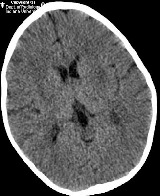
CT head: Low attenuation in the bilateral basal ganglia
MRI brain: There are small focal areas of diffusion restriction in the periventricular white matter, bilateral thalami and putamen. There is T2 prolongation affecting the basal ganglia, corpus callosum (genu and splenium), external capsules, and corona radiata.


Diagnosis: Hemolytic uremic syndrome


Key points

Most common pathogen is E. Coli O157:H7 verotoxin.
Clinical: acute renal failure, microangiopathic hemolytic anemia, thrombocytopenia, and neurologic symptoms
Neurologic complications occur in 20-50%. Symptoms include seizures, visual changes, altered consciousness and brainstem findings.
Neurologic findings most likely due to verotoxin affecting the microvascular endothelium leading to small vessel infarction and/or hemorrhage.
Patients with neurologic symptoms should undergo CT and MRI evaluation.
Treatment: fluid replacement, plasmapheresis. Avoid antibiotics which can worsen HUS.


Imaging (MRI)


Basal ganglia T2 hyper intensity is the most common finding, particularly the dorsal lateral lentiform nuclei.
Involvement of the basal ganglia is associated with a good clinical outcome.
T2 weighted hyper intensities have been reported to involved the thalamus, internal and external capsules, dorsal brainstem, corpus callosum, posterior leukoencephalopathy syndrome. Findings likely related to edema.
Diffusion restriction in the basal ganglia and thalami has been reported.
Areas of T1 hyper intensity should raise the possibility of hemorrhage.
Areas of hemorrhage are most likely to result in neurologic sequela.