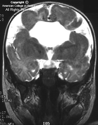

Findings
There are bilateral complete clefts in the posterior frontal parietal regions, with the lateral ventricles communicating openly with the subdural space. These clefts are lined with gray matter (polymicrogyria). The septum pellucidum is absent, and the corpus callosum is hypoplastic.
Diagnosis: Schizencephaly
Schizencephaly is a disorder of neuronal migration during embryogenesis of the brain in which clefts extend from the lateral ventricles to the pia. These clefts are lined by gray matter, may be unilateral or bilateral, and range in size from small slits to large gaps in the surrounding cerebral hemisphere. The etiology is unknown, but its frequent association with other disorders, including heterotopic gray matter, dysplasia of the corpus callosum, and absence of the septum pellucidum, points to a common error in migration. Other associations include the presence of arachnoid cysts, mega cisterna magna, and calcifications (best seen on CT). Clinically, epilepsy has been reported in 50-80% of cases of schizencephaly, more commonly in association with unilateral than with bilateral clefts; but the seizures tend to begin earlier and have a worse outcome with bilateral defects. The most common clinical finding is of asymmetrical muscle tone, which ranges from asymptomatic to paraparesis; increasing severity is associated with frontal location and bilaterality of clefts. Delay in motor skills, language deficits (more common in temporal defects), and hydrocephalus are also frequently encountered. MR is the modality of choice for diagnosis and for differentiation from porencephaly. The key finding is that the cleft is lined by gray matter, which is pathognomonic for schizencephaly. Other disorders of migration that may demonstrate enlarged or joined ventricles include lissencephaly and holoprosencephaly.
Nessun commento:
Posta un commento