venerdì 27 novembre 2009

Intraosseous cavernous hemangioma of the left maxillary bone


Findings

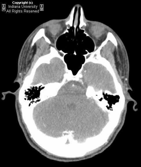
Noncontrast orbital CT demonstrates a mass arising from the left maxillary bone with internal bony trabeculations in a “honeycomb,” “sunburst,” or “spokewheel” pattern typical for an intraosseous cavernous hemangioma.
This pattern of trabeculation on CT translates into a network of linear hypointense T1 and T2 signal with interspersed areas of enhancement on the T1 post gadolinium images characteristic of this entity.


Diagnosis: Intraosseous cavernous hemangioma of the left maxillary bone


Intraosseous cavernous hemangiomas are benign neoplasms which comprise only up to 1% of all osseous tumors and 7-10% of skull tumors. These are most frequently seen in the frontal and parietal bones with the orbital and maxillofacial bones infrequently involved. These tumors are slow growing and are generally asymptomatic unless they cause mass effect on sensitive structures such as within the intraorbital compartment. In symptomatic cases, en bloc resection is generally the treatment of choice. Preoperative angiography with potential embolization may be helpful as these tumors can bleed profusely.

Clinical differential diagnosis includes fibrous dysplasia, osteoma, dermoid cyst, meningioma, eosinophilic granuloma, Lagerhan cell histiocytosis, and metastatic disease. On imaging, these tumors are well-defined and have characteristic internal trabeculations and enhancement features described as a “honeycomb,” “sunburst,” or “spokewheel” pattern. Malignant degeneration is rare.

mercoledì 25 novembre 2009

Petrous apex chondrosarcoma


Findings

The head CT (bone windows) demonstrates a region of bony destruction extending from the right petro-occipital fissure into the carotid canal. The axial T2 images demonstrate a hyper intense, extra-axial, right petrous apex mass that extends from the petro-occipital fissure to the cavernous sinus. The margins of the mass are lobulated, and it encases the cavernous internal carotid artery. The axial T1 contrast-enhanced images demonstrate intense enhancement of the mass.


Diagnosis: Petrous apex chondrosarcoma


Key points

Classic MR imaging appearance of petrous apex chondrosarcoma is a mass located at the petro-occipital fissure with high T2 signal intensity that heterogeneously enhances.
The CT shows chondroid mineralization in 50%.
Invasive bone changes at the petro-occipital fissure strongly favors the diagnosis. Greater than 50% will have associated bone destruction.
2/3 located at the petro-occipital fissure, 1/3 located at the anterior basis sphenoid
Usually has lobulated margins
High T2 signal, low to intermediate T1 signal
Heterogeneous enhancement with contrast; whorls of enhancement within tumor matrix are often seen
Often displaces or encases the ICA
Classic presentation is a CN 6 nerve palsy. Other CN palsies can occur less commonly (3, 5, 7, 8—3, 4, and 6 are possible with cavernous sinus invasion)


Differential diagnosis
- Metastatic tumor
- Chondrosarcoma
- Plasmacytoma
- Nasopharyngeal cancer
- Chordoma
- Cholesteatoma
- Calcified meningioma
- Chondromyxoid fibroma

Distinguishing characteristics from other entities in the differential:
- Metastatic tumor – Can have a similar appearance; breast cancer and prostate cancer are two of the most common tumors to metastasize to the petrous apex
- Plasmacytoma - Usually intermediate T1 and T2 signal; usually more midline
- Chordoma - Similar appearance to chondrosarcoma but often see tumor "thumb" indenting the anterior pons; often midline
- Cholesteatoma - Does not enhance but has an otherwise similar appearance
- Calcified meningioma – Not typically destructive; low to intermediate T2 signal
- Chondromyxoid fibroma - Similar MR appearance to chondrosarcoma; areas of ground glass density can be seen on CT

giovedì 19 novembre 2009

Persistent trigeminal artery (PTA)


Findings

There is an aberrant artery arising from the cavernous segment of the right internal carotid artery, coursing posterior and medial to terminate in the basilar artery. The basilar artery is diminutive below the level of this vessel and is normal in caliber above it.


Differential diagnosis

Variant caroticobasilar anastomosis
- Persistent trigeminal artery (PTA)
- Persistent hypoglossal artery (PHA)
- Persistent otic artery (POA)
- Fetal origin of the posterior cerebral artery (FOPCA)


Diagnosis: Persistent trigeminal artery


Key points

Persistent right trigeminal artery (PTA) is the most common (0.1 - 0.2% of angiograms) of a group of persistent embryonal carotid-basilar artery anastomoses.
Multiple transient connections occur consecutively in fetal life between the carotid and vertebrobasilar circulations (named according to the cranial nerve they parallel):

- Proatlantal intersegmental artery – connects cervical ICA (C2-C3) to the vertebral artery between C1 and the occipital bone
- Persistent hypoglossal artery – cervical ICA (C1-C2) and proximal (caudal) basilar artery; 2nd most common
- Persistent otic artery – petrosal ICA to proximal basilar artery via the internal acoustic meatus; very rare; vertebral arteries may be absent or hypoplastic
- Persistent trigeminal artery – cavernous ICA to distal basilar; may see enlargement of the basilar above the level of this artery; supplies basilar before the vertebral and posterior communicating arteries develop.

Clinical significance: 25% prevalence of other vascular abnormalities (e.g. aneurysms)
Usually asymptomatic but may present with mass effect on adjacent structures like the pituitary and cranial nerves.
No treatment needed if asymptomatic.

martedì 17 novembre 2009

Mondini malformation


Findings

Non-contrast axial CT images through the temporal bones demonstrate incomplete partitioning of the left cochlea and nonvisualization of the modiolus resulting in confluency between the middle and apical segments. Compare this to the right cochlea, which is normal. There is also abnormal dilitation of the left vestibular aqueduct.


Diagnosis: Mondini malformation


Case points

Mondini malformation occurs when there is abnormal development of the cochlea resulting in less than 2.5 turns and incomplete formation of the modiolus.
It is a cause of sensorineural hearing loss.


Discussion

Mondini malformation refers to an anomaly of the cochlea in which the modiolus fails to develop properly in the seventh week of fetal life, resulting in a cochlea with only 1.5 turns instead of the normal 2.5 turns. In addition, the interscalar septum between the middle and apical segments fails to form leading to a confluent, sac-like cochlea. There is an association with an enlarged vestibular aqueduct and anomalies of the vestibules and semicircular canals. In some cases, Mondini malformation may also be associated with Pendred Syndrome (bilateral sensorineural hearing loss and goiter)

Patients present with gradual or sudden sensorineural hearing loss with preservation of high-frequency hearing because the basal turn of the cochlea is usually preserved. These patients are also at increased risk for developing meningitis or perilymphatic fistula.

Treatment revolves around clinical presentation. Cochlear implants may be used to enhance hearing while middle ear infections are treated aggressively due to the increased risk for meningitis.

venerdì 13 novembre 2009

Acrania


Findings

Figure 1 and Figure 2: Coronal and axial ultrasound images of the fetal head show an absent cranial vault and an abundant amorphous mass of brain tissue. Normal cranial vault appears as bright echogenic lines characteristic of bone which is absent here.


Diagnosis: Acrania


Acrania is a rare congenital anomaly in which the flat bones of the cranial vault are partially or completely absent with complete but abnormal development of the cerebral hemispheres. The condition is identified by the absence of the calvarium. The cerebral hemispheres are surrounded by a thin membrane. The demonstration of abundant, abnormal brain tissue on the sonogram helps differentiate cases of acrania from the more common anencephaly.
Acrania is thought to occur because of abnormal migration of mesenchymal tissue, which normally covers the cerebral hemispheres. This faulty migration results in the faulty formation of the cranial bones, muscles, and dura mater. Ossification of the fetal cranium begins and accelerates after 9 weeks. The important ultrasound feature is an absent cranium, which allows diagnosis from 11 weeks onward. At 11-14 weeks gestation, the majority of cranial ossification is in the lateral aspects of the frontal bones and lower parietal bones, and no vault ossification is visible in the midline on a perfect midsagittal image. Misdiagnosis may occur if only midsagittal views of the fetus are obtained. The absence of cranial ossification may not be noted on routine examination, and the head may appear relatively normal. It is important to look specifically for frontal bone ossification in the axial and coronal planes.
The differential diagnosis includes anencephaly, large cephaloceles, hypophosphatasia and osteogenesis imperfecta. Anencephaly and acrania may commonly be confused with each other early in the pregnancy. Typically there is a progression in utero from a relatively normal appearing brain, to an amorphous brain mass, to no recognizable brain tissue which is the key to diagnosis of anencephaly in the second and third trimesters. In the first trimester, the brain of affected fetuses may appear relatively normal or may demonstrate varying degrees of distortion which can lead to confusion of accurate diagnosis. With anencephaly, the cerebral tissue is completely absent. With cephaloceles, the cranial vault can always be detected and a part of the brain is intracranial. Certain conditions such as hypophosphatasia and osteogenesis imperfecta can mimic acrania due to the lack of mineralization of the skull bones. With these conditions the intracranial anatomy is normal and the brain is surrounded by a thick layer of tissue representing soft tissues and unossified bone. Other findings such as bowing, shortening, or fractures of long bones are usually also present and aid in distinction from acrania.
Acrania is a lethal malformation. Antenatal identification allows the clinician to make appropriate and timely management decisions.

giovedì 12 novembre 2009

Submandibular sialadenitis


Findings

The left submandibular gland is hypervascular, inflamed and markedly enlarged (Figure 1 and Figure 2). There is dilatation of the submandibular duct leading to a calculus within the distal aspect of the duct (Figure 3 and Figure 4). There are no drainable fluid collections. There is injection and stranding of the overlying dermis (Figure 5). The right submandibular gland is unremarkable (Figure 2).


Diagnosis: Submandibular sialadenitis


Acute sialadenitis may be secondary to a bacterial/viral infection or an obstructing lesion such as a calculus or tumor at the floor of the mouth.
Associated conditions include HIV, sarcoidosis, Sjogren syndrome, dehydration, diabetes mellitus and immunocompromised/postoperative patients.
Imaging is often helpful to delineate the location of the calculus and the presence/absence of subsequent complications (abscess formation, osteomyelitis, etc).

Inflammation of the submandibular gland accounts for approximately 10-15% of cases of sialadenitis involving any of the major salivary glands. Risk factors for submandibular sialadenitis include immunocompromised/postoperative patients, debilitation, elderly patients, dehydration, diabetes mellitus, hypothyroidism, hypercalcemia, radiation/chemotherapy, eating disorders (bulimia, anorexia nervosa), and other concomitant medical problems (malignancy, head and neck infections). Associated conditions also include HIV, sarcoidosis, Sjogren syndrome, tuberculosis, mumps, and cat scratch disease. It is rare in pediatric patients.

Most commonly, acute sialadenitis is a result of a bacterial infection (common organisms include Staphylococcus aureus, Streptococcus, Haemophilus influenzae, and Pseudomonas). Less commonly, the infection may be related to a virus such as mumps, coxsackie virus, herpes and influenza. On the other hand, chronic sialadenitis is usually a result of salivary stasis, ductal stenosis, calculi or other obstructive lesions such as a tumor at the floor of the mouth. Sialolithiasis is most common in the submandibular gland, accounting for approximately 80% of cases. The majority of calculi are radio-opaque, vary in size, and can be single or multiple. Often, the calculus obstructs a duct, resulting in secondary inflammation of the affected salivary gland which then becomes suppurative.

Although plain film radiography can depict the majority of calculi, CT is often first-line imaging. The affected submandibular gland is enlarged, hypervascular and there may be associated cellulitis/myositis. Calculi are easily identified and described as being either distal (towards the ductal opening) or proximal (towards the submandibular hilum). Chronic sialdenitis manifests as a small, fatty gland.

Complications of sialadenitis include abscess formation, bacteremia/septicemia, osteomyelitis, cranial nerve involvement (facial nerve paralysis), and respiratory complications. Management of acute sialadenitis includes both medical (conservative) and surgical options. Antibiotics, analgesics, sialogogues, warm compresses, glandular massage and intravenous fluids are the mainstay of medical management. Surgical options include duct cannulation with subsequent removal of the calculus and complete gland excision.

mercoledì 11 novembre 2009

Bilateral parotid gland MALT lymphoma in a patient with Sjögren’s and sicca syndrome


Findings

CT Neck: Enlarged and cystic change of the bilateral parotid glands and submandibular glands without discrete enhancing mass.
MR neck: Diffuse cystic replacement and enlargement of the bilateral parotid glands (and to a lesser degree submandibular glands). No discreet enhancing parotid mass identified.

Differential diagnosis
- Non-Hodgkin's lymphoma parotid
- Benign lymphoepithelial lesions – HIV (BLL-HIV)
- Sarcoid
- Warthin tumor


Diagnosis: Bilateral parotid gland MALT lymphoma in a patient with Sjögren’s and sicca syndrome.


Key Points

Sjögren's syndrome is the second most common autoimmune disease after rheumatoid arthritis.
Sjögren's syndrome is a chronic autoimmune exocrinopathy causing salivary and lacrimal gland destruction.
Clinical: Recurrent acute episodes of glandular swelling (and dry eye, mouth, skin).
CT findings: Parotid enlargement, numerous cystic lesions, +/- punctuate calcifications
MR findings: Diffuse cystic lesions (T1 hypo, T2 hyper); heterogeneous mild enhancement of nodular parenchyma and fibrosis on T1+C, with non-enhancing cysts
Appearance of cysts helps in staging: Cysts 1-2 mm suggest Stage I or II disease. Larger cysts of > 2mm suggest Stage III, IV
Stages II-IV are readily seen on parotid US
Non-Hodgkin's lymphoma frequently complicates chronic Sjögren's parotid involvement
Bilateral parotidectomy in this patient was performed for recurrent infection / intractable pain and the surgical pathology showed bilateral MALT lymphoma