


Findings
Sagittal T1 (Figure 1): There is an isointense posterior epidural mass that extends inferiorly from the C4-C5 disc space level and displaces the spinal cord anteriorly.
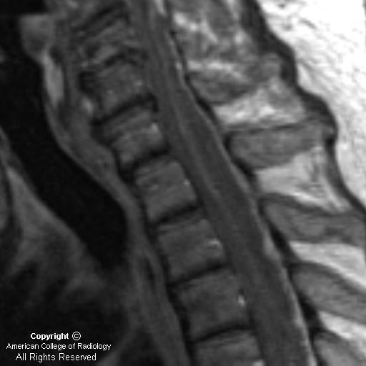
Sagittal T2 (Figure 2): The posterior epidural high-signal mass is more well-defined on this T2-weighted image.
Axial T2 (Figure 3): Image demonstrates compression of cord elements by the posterior epidural mass.
Post-contrast Sagittal T1 (Figure 4): Image demonstrates an enhancing posterior epidural collection consistent with an abscess.
Diagnosis: Spinal epidural abscess
A spinal epidural abscess (SEA) can present with nonspecific signs and symptoms, including lower back pain, lower extremity weakness, or even sepsis. Cord compression can result if an epidural abscess in the spinal canal is not promptly treated. In a patient with back pain and fever, an SEA should be considered until proven otherwise.
An SEA can result through hematogenous spread or from direct extention of adjacent discitis or osteomyelitis. Remote infections, from indwelling catheters or even urinary tract infections, may hematogenously spread to create an SEA. Alternatively, any procedure in which there is direct puncture into the spinal canal, may seed an infection that leads to the development of an epidural abscess. Additional risk factors for the development of SEA include IV drug abuse, diabetes, alcoholism, and chronic immunosuppression.
MRI with gadolinium is the test of choice in evaluating patients suspected of having an epidural abscess. Alternatively, CT myelography can be utilized in patients who have contraindications to MRI. A lumbar puncture is a relative contraindication if a spinal epidural abscess is suspected, because infectious agents may be introduced into the subarachnoid space.
MRI findings usually fall into 2 categories: 1) A soft tissue mass that is hypointense on T1-weighted images and hyperintense on T2-weighted images with diffuse homogeneous or slightly heterogeneous enhancement within the collection; 2) As the phlegmonous mass necroses, there may be a peripherally-enhancing fluid collection. Spinal epidural abscesses may be located anteriorly or posteriorly within the spinal canal. Anterior epidural abscesses are generally secondary to spread of adjacent infection from discitis or osteomyelitis. Posterior epidural abscesses may occur as a result of hematogenous spread of remote infections. Other causes of back pain that should be considered in the differential diagnosis include herniated disc, neoplasm, spinal hematoma, and transverse myelitis.
Those patients who do not present with neurological symptoms may be treated with medical therapy alone. Those with neurological compromise are treated with surgical decompression and antibiotics. Our patient underwent successful surgical decompression of his spinal epidural abscess.
Nessun commento:
Posta un commento