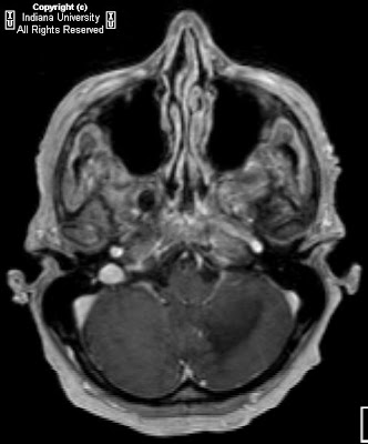


History: Woman with lung transplant and weakness.
Additional clinical information: The patient has CSF positive for JC virus and EBV virus.
Findings
Earlier CT showed an ill-defined process of the left cerebellar hemisphere, thought to represent ischemia, but was shown to progress over serial scans, making ischemic process unlikely. The MR shows T2 prolongation within the left cerebellar white matter extending across the middle cerebellar peduncle and into the left anterior pons. There is also a chronic right frontal lobe infarct.
Differential diagnosis:
- Progressive multifocal leukencephalopathy
- Encephalomalacia
- CMV encephalitis
- Lymphoma
- Toxoplasmosis
- Abscess
Diagnosis: Progressive multifocal leukencephalopathy (PML)
Key points
JC virus infection of oligodendrocytes causes demyelinating lesions
Often occurs as reactivation of latent virus in an immunosuppressed patient
Most prevalent in AIDS, leukemia, and organ transplant patients
3rd most common cause of encephalopathy in AIDS patients after toxoplasma encephalitis and HIV encephalitis
Responds to immune-strengthening treatment, such as HAART in AIDS; 8% spontaneous resolution
Clinical presentation
Insidious onset of focal symptoms, behavioral, speech, cognitive, motor, and visual impairment, over weeks
More rapid progression than AIDS dementia complex
Conjugate gaze abnormalities are common and are the initial presentation in more than 30% of patients
Radiology
Contrast-enhanced CT
- Multifocal, nonenhancing white matter hypodensities without mass effect or edema.
- Rapid change in size or number of lesions
MRI
- Hypointense T1-weighted appearance with cortical sparing; typically nonenhancing; occasionally, mild peripheral enhancement
- T2-weighted images reveals hyperintense subcortical lesions
Nessun commento:
Posta un commento