


Findings
Figure 1 and Figure 2: Sagittal T1 and T2 weighted images reveal the presence of signal in the IVC and iliac veins indicative of thrombus. Prominent epidural veins are appreciated here as well.
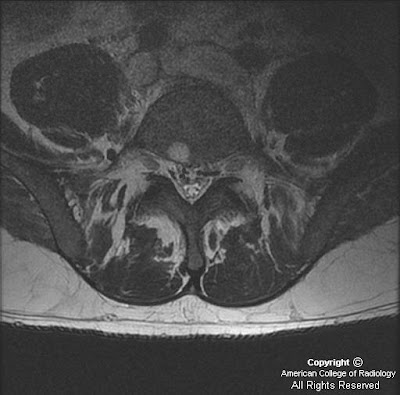
Figure 3 and Figure 4: Axial T2 weighted sequences reveal enlarged epidural veins as demonstrated by prominent flow voids in the epidural space. Note absent flow void in the IVC and iliac veins. Normal flow voids are seen in the associated arterial structures on these images.
Diagnosis: IVC thrombosis with enlarged epidural veins causing a lumbar radiculopathy
Lumbar radiculopathy and low back pain is a frequently encountered complaint in the general population. The most common etiology is structural spine disease, including disc herniation and spondylosis. However, nonstructural diseases, such as cytomegalovirus polyradiculopathy in immunocompromised individuals as well as other infections, neoplasms, and inflammatory conditions have also been shown to cause lumbar radiculopathy. Vascular causes are unusual as patients with vascular pathology tend to present with myelopathy.
Enlarged epidural veins are commonly a result of arteriovenous malformations, fistulas, and varicose veins. In the cervical spine, the epidural veins function as collateral pathways and become enlarged as they receive more blood flow when the jugular veins are compromised. Similarly, it can be inferred that in the case of an IVC obstruction or occlusion, the lumbar epidural veins become enlarged by the same mechanism; when IVC blood flow is compromised, blood is able to return to the heart through the azygos and hemiazygos veins via collateral pathways including the epidural venous system. While many epidural veins become enlarged as a result of this process, the vein implicated in causing the radiculopathy is the vein below the pedicle. This vein lies in close proximity to the existing nerve root and as the vein becomes larger, it will impinge directly upon the nerve root leading to the radiculopathy. Therefore, the best way to eliminate the radicular symptoms is by treating the underlying cause of the IVC thrombosis.
Risk factors for the development of IVC thrombosis and venous thromboembolism (VTE) include hypercoaguable states such as factor V Leiden mutation, prothrombin gene mutation, protein S deficiency, protein C deficiency, antithrombin deficiency, and dysfibrinogenemia. Acquired risk factors include a prior thrombotic event, recent major surgery, presence of a central venous catheter, trauma, immobilization, malignancy, pregnancy, oral contraceptives, heparin, myeloproliferative disorders, and antiphospholipid syndrome.
Excellent topics, I like this. I found this online that, I know Arkansas Spinal Care Conway, AR their commitment to their patients has ledthem to using the DRX spinal decompression equipment for low back pain.
RispondiEliminachiropractor conway ar
Spider veins can be unsightly and sometimes painful - usually appearing on the legs. Learn more about treatments telangiectasia, creams, and more.
RispondiEliminaveins
Nice topic. My spider vein removal team appreciates this so much. Keep sharing.
RispondiEliminaWhite Rock Laser Clinic has established a dominant position in the market by offering a wide range of services such wrinkles removal, birth marks, body hair removal, age spots removal,Wart and skin, Veins removal and much more. We have occupied the market by creating a powerful brand and large customer base through delivering reliable and healthy cosmetic treatments.
RispondiElimina