martedì 30 marzo 2010

Fahr disease


Findings

Figure 1 and Figure 2: Select axial non-contrast CT images demonstrate dense symmetric calcification in the basal ganglia (Figure 1) and dentate nuclei (Figure 2). Cortical atrophy is also seen.
Figure 3: Axial T1-weighted image demonstrates symmetric cerebellar hyperintense foci corresponding to calcification seen on CT.


Diagnosis: Fahr disease (Idiopathic familial basal ganglia calcification)


Fahr disease (Idiopathic familial basal ganglia calcification, bilateral striopallidodentate calcification, Familial cerebrovascular ferrocalcinosis) is a rare disorder characterized by idiopathic basal ganglia calcification with associated cognitive and neurobehavioral manifestations.

Calcification is found primarily in the globus pallidus, but the putamen, caudate, thalamus, cerebellum (especially dentate nucleus), corona radiata, and subcortical white matter can also be affected. There are no detectable abnormalities of calcium or phosphate metabolism. Patients develop progressive parkinsonism, dystonia, and neuropsychiatric disturbance.

There is a bimodal pattern of onset. Those affected in early adulthood may be asymptomatic in the first two decades, despite the presence of basal ganglia calcification. The disease presents with schizophreniform psychosis. A second peak of onset is seen in late middle age, when patients present with subcortical dementia or Parkinsonian symptoms, which are permanent and progressive. Paroxysmal dystonic choreoathetosis and seizures are common. Eventually patients develop symmetrical spastic paralysis progressing to a decerebrate state.

The disease process involves the deposition of calcium in the walls of the capillaries and larger arteries and veins. Other elements, including magnesium, zinc, aluminum, and iron have also been found deposited in the vessels. No definitive treatment is available.

In studying a three-generation family with an autosomal dominant form of the disease, Geschwind et al, established that the chromosomal locus, IBGC1, lies on chromosome 14, and found that this form of the disease demonstrates genetic anticipation. Autosomal recessive inheritance has also been documented. The disease demonstrates variable expressivity.

CT images demonstrate bilateral, symmetric calcification in the globus pallidus, cerebellum, and white matter. On T1-weighted MR images calcifications are hyperintense, while on T2-weighted and FLAIR images, calcification may be hypo- or hyperintense. T2 hyperintense regions in the white matter, which do not correspond to calcification can also be seen. This finding may reflect progressive inflammation.

Differential diagnosis for inherited and acquired basal ganglia calcification
Postinflammatory causes:
- Tuberculosis
- Toxoplasmosis
- Cystercercosis
- Congenital HIV

Endocrine causes:
- Hyperparathyroidism
- Hypoparathyroidism
- Pseudohypoparathyroidism
- Hypothyroidism

Congenital causes:
- Tuberous sclerosis
- Down syndrome
- MELAS
- Neurofibromatosis

Toxic causes:
- Exposure to carbon monoxide
- Chemotherapy
- Radiation therapy
- Lead intoxication

In addition, incidental basal ganglia calcification is seen frequently on CT imaging in patients over the age of 50.

venerdì 26 marzo 2010

Mechanical thrombectomy of the MCA


Findings

Figure 1: CT angiography of the head demonstrates a left MCA occlusion at the distal M1 area. There is visualization of distal vessels with filling by collaterals.

Figure 2, Figure 3, Figure 4, and Figure 5: CT perfusion images demonstrate increased transit time and poor flow in the left MCA territory. There is poor flow in the right posteroparietal region. There is generally preserved blood volume except for decreased volume in the left posterior watershed area. These findings are compatible with ischemia with some areas of infarct and fairly large area of preserved viability.

Diagnostic/therapeutic cerebral angiogram findings: Digital angiography confirms that the left M1 segment is occluded at its mid-portion (Figure 6). Subsequently, the Merci catheter is deployed in the region of the thrombus (Figure 7). Following Merci thrombectomy, there is recanalization of the left M1 segment of the middle cerebral artery (Figure 8).


Diagnosis: Mechanical thrombectomy of the MCA


Catheter-directed thrombectomy systems can be successfully applied towards salvaging areas of reversible ischemia detected on cerebral perfusion imaging.
The MERCI catheter thrombectomy technique involves insinuating the catheter’s coiled tip beyond the thrombus and then retracting the catheter, thereby extracting the clot material.

Until recent years, intravenous recombinant tissue plasminogen activator (TPA) administered within three hours following symptom onset was the only FDA approved treatment for acute stroke. However, mechanical thrombectomy has since emerged as an effective treatment option for acute cerebral ischemia. Indeed, satisfactory results have been achieved with mechanical thrombectomy in some patients that present beyond the time period for intravenous TPA administration has passed, up to about 8 hours. Specifically recanalization rates of nearly 70% have been reported using mechanical thrombectomy.

Various catheter-based devices and techniques have now been devised for mechanical thrombectomy in the cerebral arteries. Some of these include the use of a snare, the alligator retrieval system, the Phenox clot retriever, the Merci catheter, among others.

The Merci catheter thrombectomy technique consists of obtaining femoral artery access, guidewire insertion, and advancement of the catheter to the site of occlusion. Subsequently, the balloon is inflated and the coiled tip of the catheter is passed across the clot and engaged under angiography. The mesh helps trap thrombus material. Clot is retracted into the balloon guide catheter and out of the body. Finally, the balloon is deflated, thereby restoring blood flow.

The main complication of mechanical thrombectomy is intracranial hemorrhage, which occurs in an estimated 5 to 10% of patients.

martedì 23 marzo 2010

Focal cortical dysplasia, Taylor type


Findings

The MRI of the brain shows T2 and FLAIR hyper intense arrow-shaped left frontal white matter lesion pointing toward the frontal horn of the left lateral ventricle. There is no abnormal enhancement. There is mild hypo intensity on T1 sequences.

Differential diagnosis:
- Focal cortical dysplasia
- Gliosis
- Glial cell tumor
- Physiologic margination of white matter neurons


Diagnosis: Focal cortical dysplasia, Taylor type


Key points

Focal cortical dysplasia is considered to be in the spectrum of migrational disorders of the brain, and often manifests in the form of seizures, developmental delay and focal neurologic deficits. Focal cortical dysplasia is thought to represent the cause of between 5% and 25% of patients with focal epilepsy. This entity typically manifests in the first years of life. Antiepileptic medications are the first line of therapy, and no particular medications have been found to be more or less effective for focal cortical dysplasia. After two failed rounds of antiepileptic regimens, surgical interventions may be considered.


Radiology

MRI is the examination of choice for identifying focal cortical dysplasia. The typical appearance of the Taylor type of this disorder is that of T2/FLAIR hyper intensity within the sub cortical white matter, tapering toward the lateral ventricle. The most typical location of this finding is unilaterally within the frontal lobes. The lesions do not enhance.

TANTI AUGURI PROFESSO'

Happy Birthday to my great friend and teacher, Eytan Raz M.D. !!!

mercoledì 17 marzo 2010

Subacute Sclerosing Panencephalitis (SSPE)


History: 18-year-old male with 3 months of personality change, gradual decrease in responsiveness, rigidity and abnormal EEG findings (the patient is HIV negative and does not report a recent viral or respiratory infection or vaccination).


Findings

Figure 1: T2-weighted axial image demonstrates multiple areas of hyperintense signal in the bilateral subcortical and deep white matter.
Figure 2: T2-weighted FLAIR axial image demonstrates areas of hyperintense signal in the posterior limb of the right internal capsule (blue arrow) and right aspect of the splenium of the corpus callosum (yellow arrow).
Figure 3: T1-weighted FLAIR post contrast image demonstrates no areas of abnormal enhancement.
Figure 4: There is no restricted diffusion.


Diagnosis: Subacute Sclerosing Panencephalitis (SSPE)


SSPE is a chronic progressive encephalitis that can very rarely occur in people 2-10 years following infection with the measles virus. Early stages are characterized by personality, behavioral and intellectual changes. There is then further neuropsychiatric deterioration progressing to ataxia, chorea, dystonic rigidity, seizures, myoclonus, optic atrophy and cortical blindness. The terminal stages include unresponsiveness, autonomic dysfunction, coma and death within 1 to 3 years of the onset of symptoms.

The white matter is predominantly affected with patchy demyelination and initial sparing of the subcortical U fibers. In the first stage of the disease, T2WI/FLAIR typically demonstrates asymmetrical gray and adjacent subcortical white matter hyperintensity in the parieto-occipital regions. Enhancement and mass effect may occur early in the disease. The basal ganglia (particularly the putamen), cerebellum, pons and frontal white matter may also be involved. In the second stage of the disease, cortical/subcortical lesions regress and T2WI/FLAIR periventricular hyperintensity develop. In the terminal stage, periventricular white matter disease progresses and profound parenchymal loss occurs.

Diagnosis is made by characteristic CSF and EEG findings. In SSPE, CSF will typically have normal cellular components, glucose and total protein, but markedly elevated values of gammaglobulin and anti-measles antibodies. Typically, serum anti-measles antibody titers are also grossly elevated. A characteristic “burst-suppression” pattern on EEG is nearly always seen.

Treatment is the immunomodulator interferon and antivirals ribavirin and inosine pranobex which, if given early and throughout the patient’s life, may limit progression of disease.

martedì 16 marzo 2010

Progressive multifocal leukencephalopathy (PML)


History: Woman with lung transplant and weakness.
Additional clinical information: The patient has CSF positive for JC virus and EBV virus.


Findings

Earlier CT showed an ill-defined process of the left cerebellar hemisphere, thought to represent ischemia, but was shown to progress over serial scans, making ischemic process unlikely. The MR shows T2 prolongation within the left cerebellar white matter extending across the middle cerebellar peduncle and into the left anterior pons. There is also a chronic right frontal lobe infarct.

Differential diagnosis:
- Progressive multifocal leukencephalopathy
- Encephalomalacia
- CMV encephalitis
- Lymphoma
- Toxoplasmosis
- Abscess


Diagnosis: Progressive multifocal leukencephalopathy (PML)


Key points

JC virus infection of oligodendrocytes causes demyelinating lesions
Often occurs as reactivation of latent virus in an immunosuppressed patient
Most prevalent in AIDS, leukemia, and organ transplant patients
3rd most common cause of encephalopathy in AIDS patients after toxoplasma encephalitis and HIV encephalitis
Responds to immune-strengthening treatment, such as HAART in AIDS; 8% spontaneous resolution


Clinical presentation

Insidious onset of focal symptoms, behavioral, speech, cognitive, motor, and visual impairment, over weeks
More rapid progression than AIDS dementia complex
Conjugate gaze abnormalities are common and are the initial presentation in more than 30% of patients


Radiology

Contrast-enhanced CT
- Multifocal, nonenhancing white matter hypodensities without mass effect or edema.
- Rapid change in size or number of lesions

MRI
- Hypointense T1-weighted appearance with cortical sparing; typically nonenhancing; occasionally, mild peripheral enhancement
- T2-weighted images reveals hyperintense subcortical lesions

martedì 9 marzo 2010

Hydrancephaly


Findings

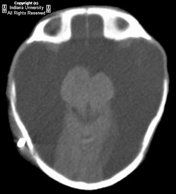
There is near complete absence of the cerebrum, with small amount of residual occipital lobe. Thalami, cerebellum, and brainstem are present.

Differential diagnosis:
- Hydranencephaly
- Hydrocephalus
- Bilateral schizencephaly
- Alobar holoprosencephaly


Diagnosis: Hydrancephaly


Key points

Hydranencephaly - In utero destruction of cerebral parenchyma with intact falx and preservation of posterior fossa structures.
Cerebrum replaced with CSF.
Caused by in utero occlusion of bilateral supraclinoid internal carotid arteries. Etiology unclear (hereditary thrombophilic states, infection, maternal irradiation/toxin exposure, twin-twin transfusion, intrauterine anoxia).
Rarely unilateral
Occurs approximately <1:10000 births, greatest incidence in teenage mothers.
Clinically, present with macrocephaly, developmental delay, irritability, hyperreflexia, seizures.
Often seen with prenatal ultrasound (anechoic cranial vault).
On CT and MR (best characterized with MR) – CSF attenuation/signal intensity replacing the supratentorial brain parenchyma with sparing of thalami, brain stem, cerebellum, and choroid plexus.
Falx is intact (distinguishes from holoprosencephaly).
No thin rim of cerebral parenchyma (distinguishes from severe hydrocephalus).
Prognosis is poor – usually death in infancy.
Treatment is supportive care, shunting to decrease head growth.

sabato 6 marzo 2010

Colloid cyst


Findings

Seven mm hyperdense round lesion in the third ventricle with mild ventricular enlargement for age. Bone window images showed that the lesion was not calcified. MR shows bright signal on T1 consistent with increased protein. T2 demonstrates isointense to brain signal. T1 post contrast shows some peripheral enhancement without clear enhancement of the lesion.

Differential diagnosis for third ventricular masses:
- Colloid cyst
- Subependymal giant cell astrocytoma
- Craniopharyngioma
- Ependymoma
- Meningioma
- Choroid plexus papilloma
- Vascular lesion
- Intraventricular neurocysticercosis


Diagnosis: Colloid cyst


Key points


Colloid cysts are rare brain lesions that account for approximately 1-3% of primary intracranial tumors. The typical location is in the anterior superior third ventricle. Rare cases have been reported in the lateral ventricles, fourth ventricle, and even outside the ventricular system. Diagnoses usually occurs incidentally in adult patients. Very rarely diagnosed in children. Colloid cysts originate during the development of the nervous system from ectopic endodermal elements which migrate into the velum interpositum. These lesions are lined by cuboidal or stratified columnar epithelium and contain proteinaceous components. This lining accounts for the enhancement that is sometimes seen as in this case. The proteinaceous material is derived from the breakdown products and secretions of the epithelial cells.

In many patients, colloid cysts remain clinically silent. They enlarge slowly or stop growing, allowing a balance to develop between the production and absorption of CSF. Even with this slow growth some patients experience signs and symptoms of hydrocephalus due to the obstruction of the foramina of Monro. They experience headache, memory deficits, and nausea/vomiting. The headache is often positional because the foramina of Monro may only become obstructed in certain positions as the cyst is often somewhat mobile. Cases of sudden death have been reported. Although these lesions are benign, they are often surgically managed since the complication of obstructive hydrocephalus may be life threatening.

On CT scans, most are hyperdense to the brain although they are not calcified. Occasionally, they may be hypodense or isodense. The appearance on MRI is variable as well. Most lesions have high signal intensity on T1 and low signal intensity on T2, though they may vary from high to low intensity on both T1 and T2. Thin rim enhancement occasionally is seen as in this case.

mercoledì 3 marzo 2010

Adult medulloblastoma


Findings

Figure 1 and Figure 2: Axial noncontrast CT. There is a 5.3cm x 4.2 cm hyperdense, lobulated mass in the right cerebellum. There is mass effect upon the fourth ventricle with midline shift.
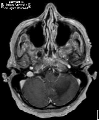
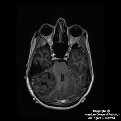
Figure 3: Axial FLAIR. A large iso intense 4.5 cm x 5.0cm x 3.4 cm mass is seen in the right lateral cerebellum.
Figure 4: Axial SPGR. A large hypointense 4.5 cm x 5.0cm x 3.4 cm mass is seen in the right lateral cerebellum.
Figure 5: Axial T2. A large 4.5 cm x 5.0cm x 3.4 cm soft tissue signal heterogeneous mass is seen in the right lateral cerebellum with foci of high T2 signal. T2 signal hyperintensity suggests multiple small cystic regions. There is moderate right to left midline shift with mass effect on the fourth ventricle.
Figure 6: Axial T1 post. A large hypointense 4.5 cm x 5.0cm x 3.4 cm mass is seen in the right lateral cerebellum. There is minimal central enhancement with gadolinium.
Figure 7: Coronal T1 post. A large heterogeneous 4.5 cm x 5.0cm x 3.4 cm mass is seen in the right lateral cerebellum. There is minimal central enhancement with gadolinium.
Figure 8: Sagittal T1 post. A large heterogeneous 4.5 cm x 5.0cm x 3.4 cm mass is seen in the right lateral cerebellum. There is minimal central enhancement with gadolinium.


Diagnosis: Adult Medulloblastoma


Medulloblastoma is a highly malignant neuroepithelial tumor of the posterior fossa that arises from transient undifferentiated neuro-epithelial cells of the developing cerebellum. Adult medulloblastoma is an infrequent tumor representing 1% of CNS tumors and 6% of posterior fossa tumors of adults.

Headache, vomiting, ataxia, and visual deterioration are common presenting symptoms. Hemiataxia is a frequent finding in adults because of the hemispheric location of most tumors. Gait ataxia, papilledema, nystagmus, facial palsy, and neck stiffness are also common. Most (75%) patients have symptoms for less than 3 months and without treatment, medulloblastoma causes death within a few months after presentation.

A well defined homogenous vermian mass is the most common presentation of medulloblastoma in children. By comparison, the most common presentation of adult medulloblastoma is that of a poorly defined, heterogeneous hemispheric tumor with evidence of cystic and necrotic degeneration. Approximately half of adult medulloblastomas originate in a cerebellar hemisphere, are hyperdense on unenhanced CT, and show minimal enhancement after injection of contrast medium. This is most likely because of the higher prevalence of desmoplastic lesions in adults. The characteristic hyperattenuation on unenhanced CT scans reflects the high nuclear-cytoplasmic ratio seen at histologic analysis.

The tumor typically appears heterogeneous on images, findings that are related to cyst formation, hemorrhage, and calcification and that are even more pronounced with magnetic resonance (MR) imaging. As in adults, medulloblastoma has a variable MR appearance in children, On T1-weighted images, tumors are hypo- or isointense relative to cortex and on T2-weighted images, they are hypo-, iso-, or hyperintense. Evidence of leptomeningeal metastatic spread is present in 33% of all cases at the time of diagnosis and is well evaluated with contrast-enhanced MR imaging of the brain and the spine.

Although conventional MR imaging is helpful in distinguishing among infratentorial brain tumors, newer techniques, such as diffusion-weighted imaging (DWI) further faciliate this distinction. DWI provides information that cannot be obtained with conventional MRI. DWI evaluates the microscopic rate of water diffusion within tissues. It can identify different tumor types and delineate their boundaries with normal tissue. For instance, medulloblastoma will have increased signal intensity or restricted diffusion on DWI. In contrast, pilocytic astrocytoma shows no restricted diffusion on DWI.

The differential diagnosis for a posterior fossa mass include metastasis, hemangioblastoma, astrocytoma, lymphoma, and dysplastic cerebellar gangliocytoma (Lhermitte-Duclos disease). Lateral tumors in adults often appear extraaxial and may simulate meningiomas of the cerebellopontine angle or tentorium, acoustic neuromas, ependymomas, glomus jugulare tumors or metastases, solid hemangioblastoma and choroid plexus papilloma. Metastasis is the most common cerebellar mass in the adult population and hemangioblastoma is the most common primary neoplasm of the cerebellum in adults.

Surgical resection, radiation therapy, and chemotherapy have substantially lowered the mortality associated with this tumor, with 5-year survival rates now commonly well above 50%. The practice of using staging data developed for childhood medulloblastoma to determine risk for recurrence and poorer prognosis may not be valid in the adult population. The current standard of care remains surgical resection of the primary neoplasm followed by craniospinal irradiation. The optimal use of adjuvant chemotherapy remains unclear, but may be best reserved for patients with CNS or extraneural metastasis at diagnosis.

lunedì 1 marzo 2010

Pelizaeus-Merzbacher disease


Findings

There is diffuse T2 prolongation within subcortical, deep, and periventricular cerebral and cerebellar white matter with involvement of internal capsules and corpus callosum. There is also involvement of cerebellar peduncles. No enhancement or diffusion restriction.

Differential diagnosis:
- Pelizaeus-Merzbacher
- Metachromatic leukodystrophy
- Adrenoleukodystrophy
- van der Knaap leukoencephalopathy
- Canavan disease


Diagnosis: Pelizaeus-Merzbacher


Pelizaeus-Merzbacher is a rare leukodystrophy which results in abnormal myelin production. The disease is related to the proteolipid protein 1 (PLP1) gene on the X chromosome. The products of this gene constitute about 50% of the mass of CNS white matter and are believed to serve an important structural function in compact myelin. The defect is most commonly a duplication of genetic material which is deleterious to oligodendrocytes.

The frequency is not certain but is conservatively thought to be more than 1 in 500,000. Clinical signs usually include some combination of nystagmus, stridor, spastic quadriparesis, hypotonia, cognitive impairment, ataxia, and tremor. Patients tend to be normocephalic. Nerve conduction tests typically normal (as opposed to most other leukodystrophies). Diagnosis involves clinical history and physical examination, MRI of the brain, and molecular diagnostic tests. Because of the heterogeneity of genetic defects and since females tend to be heterozygous, there is varying severity and clinical course.

No specific cure or treatment for Pelizaeus-Merzbacher disease is known. Medical care is currently limited to supportive care and includes tracheostomy, physical therapy, orthotics, and antispasticity agents, including intrathecal baclofen.


Radiology

There is lack of myelin maturation on T2WI (i.e. white matter is brighter than it should be) and there is minimal myelin maturation on T1WI (i.e. white matter is darker than it should be). Changes may be difficult to identify under 1 year of age due to the normal lack of myelination. There is diffuse involvement without predilection for a specific distribution or sparing of sub cortical U-fibers. There is no enhancement (in contrast to Alexander or X-linked adrenoleukodystrophy). There is no telltale spectroscopy finding (in contrast to Canavan disease which has elevated NAA). There is no diffusion restriction (in contrast to adrenoleukodystrophy). There is no volume expansion of the white matter (in contrast to van der Knaap leukoencephalopathies).