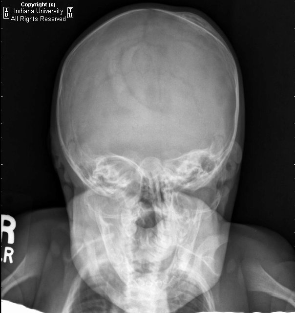
Findings
Lenticular left parietal calvarial mass-like lesion with peripheral sclerosis/calcification and central lucency. The lesion does not cross suture lines.
Differential diagnosis:
- Calcified cephalohematoma
- Caput succedaneum
- Subgaleal hematoma
- Subperiosteal osteoid osteoma
- Leptomeningeal cyst
Diagnosis: Subperiosteal cephalohematoma.
Cephalhematomas are subperiosteal hematomas that frequently occur after using instruments (forceps) during delivery. They are not associated with skull fractures and are limited by suture lines. They typically occur in the parietal region and may not appear until 2-3 days after birth. Treatment is usually not necessary. Complications include infection. Caput succedaneum is a subcutaneous hemorrhage that occurs after vaginal delivery, does cross suture lines and requires no intervention. A subgaleal hemorrhage covers a much larger area than a cephalohematoma and is potentially life threatening. It usually occurs from rupture of emissary veins in the subaponeurotic space, often secondary to vacuum suctioning.
Radiologic overview
Often seen as soft tissue subperiosteal elevation on conventional radiography. Frequency of location: parietal > occipital > frontal. The outer border may calcify. The skull at the hematoma site my remain thickened for years. It does not cross suture lines as it is bounded by the periosteum.
Nessun commento:
Posta un commento