

Findings
Figure 1: Unenhanced CT shows a heterogeneous attenuation mass with cystic spaces in the posterior fossa.
Figure 2: Intense enhancement is noted after contrast administration.
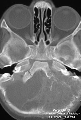
Figure 3: Bone window section showing permeative destruction of the left occipital bone.
Figure 4: Axial T1 weighted image showing extra- axial mass with multiple cystic spaces.
Figure 5: Coronal T2 image showing the extra-axial origin clearly with mass effect on the cerebellum. The cystic spaces appear hyperintense.
Figure 6: Axial T1 weighted image showing intense enhancement of the mass.
Diagnosis: Ewing sarcoma of the occipital bone
Ewing sarcoma is a small round-cell tumor arising from mesenchymal cells. These tumors affect children and young adults in the age group of 5-15 years. The long bones, flat bones like the scapula and the vertebrae are the most common sites. Primary Ewing sarcoma affecting the calvarium is extremely rare, making just 1% of the cases. In the skull, the tumor more often arises from the frontal and parietal bones and less common locations include ethmoid, temporal and occipital bones.
CT scans (bone window) reveal poorly marginated permeative destructive lesion involving both inner and outer tables of the skull. The "onion peel" appearance typical of Ewing sarcoma in long bones is not seen commonly in the calvarium. The extra-dural soft tissue shows intense enhancement on contrast administration.
MR imaging provides better soft tissue delineation of these tumors. The extra dural soft tissue appears hypointense on T1 weighted images while the cystic and necrotic areas appear hyperintense on T2 weighted images. Good contrast enhancement is noted.
The differential diagnosis should include rhabdomyosarcoma, metastatic neuroblastoma and lymphomas.
Treatment is surgery followed by chemotherapy and radiation.
Nessun commento:
Posta un commento