lunedì 29 novembre 2010

Intraventricular oligodendroglioma


Findings:

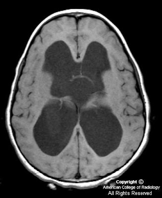
Figure 1: Non-contrast CT shows a large rounded mass in the lateral ventricles with intermediate density and large foci of calcification within it. This is causing significant hydrocephalus with enlargement of the third ventricle.


Intraventricular oligodendroglioma


Oligodentrogliomas are well differentiated, slowly growing but diffusely infiltrating cortical and subcortical tumors. Although most of them involve the frontal and frontotemporal cortex, a small proportion of them are seen in the ventricular walls (1-10%), cerebellum and exceedingly rarely within the brainstem, spinal cord and leptomeninges. No age is exempt and the peak incidence is in the 4th or 5th decades. It usually have a long standing history of symptoms and the most common being seizures and headaches.

Intraventricular oligodentrogliomas can present with hydrocephalus. Pathologically, they are well defined, grayish-pink soft unencapsulated mass and calcification is extremely common. Focal cystic degeneration and hemorrhage are frequent findings. CSF seeding is uncommon. Histopathologically, these are moderately cellular tumors with occasional mitosis. Perinuclear halos or the "fried egg artifact" is a distinctive feature of oligodentroglioma. Majority of the "intraventricular oligodentrogliomas" described in the literature are central neurocytomas and immunohistochemistry helps to distinguish them.

On CT, oligodentrogliomas usually have mixed density with nodular, clumped or linear tumoral calcification seen in majority (50-90%). Cystic degeneration is common. Intratumoral hemorrhage and edema is uncommon. The hemispheric lesions may expand, remodel or erode the calvarium. Enhancement is variable. On MR, oligodentrogliomas are typically heterogeneously hypointense or isointense to grey matter on T1 and heterogeneously hyperintense on T2 and FLAIR due to calcification, cystic change and hemorrhage. Nearly 50% show heterogeneous enhancement following contrast administration. Areas of calcification and hemorrhage demonstrate "blooming" on gradient echo MR sequences. No diffusion restriction on DWI.

Surgical resection is the primary treatment of choice. Radiation therapy and chemotherapy is reserved for recurrent tumors. Local recurrence is common and hence regular surveillance is recommended.

venerdì 26 novembre 2010

Ewing sarcoma of the occipital bone


Findings

Figure 1: Unenhanced CT shows a heterogeneous attenuation mass with cystic spaces in the posterior fossa.
Figure 2: Intense enhancement is noted after contrast administration.
Figure 3: Bone window section showing permeative destruction of the left occipital bone.
Figure 4: Axial T1 weighted image showing extra- axial mass with multiple cystic spaces.
Figure 5: Coronal T2 image showing the extra-axial origin clearly with mass effect on the cerebellum. The cystic spaces appear hyperintense.
Figure 6: Axial T1 weighted image showing intense enhancement of the mass.


Diagnosis: Ewing sarcoma of the occipital bone


Ewing sarcoma is a small round-cell tumor arising from mesenchymal cells. These tumors affect children and young adults in the age group of 5-15 years. The long bones, flat bones like the scapula and the vertebrae are the most common sites. Primary Ewing sarcoma affecting the calvarium is extremely rare, making just 1% of the cases. In the skull, the tumor more often arises from the frontal and parietal bones and less common locations include ethmoid, temporal and occipital bones.

CT scans (bone window) reveal poorly marginated permeative destructive lesion involving both inner and outer tables of the skull. The "onion peel" appearance typical of Ewing sarcoma in long bones is not seen commonly in the calvarium. The extra-dural soft tissue shows intense enhancement on contrast administration.

MR imaging provides better soft tissue delineation of these tumors. The extra dural soft tissue appears hypointense on T1 weighted images while the cystic and necrotic areas appear hyperintense on T2 weighted images. Good contrast enhancement is noted.
The differential diagnosis should include rhabdomyosarcoma, metastatic neuroblastoma and lymphomas.

Treatment is surgery followed by chemotherapy and radiation.

lunedì 22 novembre 2010

Suprasellar arachnoid cyst


Findings:

Figure 1, Figure 2 and Figure 3 show severe hydrocephalus involving the lateral and third ventricles.
Figure 4, Figure 5, Figure 6, show a suprasellar mass lesion which follows CSF signal on all sequences, including FLAIR.


Diagnosis: Suprasellar arachnoid cyst


Arachnoid cyst is the most common congenital lesion of the brain. It is typically an incidental imaging finding and is rarely symptomatic. However, in this case the size and location of the lesion in the suprasellar cistern resulted in severe obstructive hydrocephalus and precocious puberty. Both of these complications resolved following surgical drainage of the cyst.

Arachnoid cysts arise from a splitting of the arachnoid membrane with formation of a cyst wall consisting of fibrous connective tissue. There is no epithelial lining in the wall. Expansion occurs following trapping of cerebral spinal fluid through defects in the cyst wall. Arachnoid cysts occur most commonly in the middle cranial fossa and have a 4-to-1 male-to-female ratio. Even large cysts tend to be asymptomatic. Associated clinical features in symptomatic patients include headache, calvarial bulging, intracranial hypertension, craniomegaly, developmental delay, visual loss, precocious puberty, and seizures. Treatment of arachnoid cysts is not recommended by many unless there is a clear cause and effect relationship between the cyst and symptoms as shown in this case.

MRI is the preferred diagnostic modality for arachnoid cysts because of its ability to demonstrate the location, extent and relationship of the cyst to surrounding neurologic structures. Lesions typically have the signal intensity of CSF on all sequences, do not enhance and do not demonstrate restricted diffusion. The most important differential diagnostic consideration is between arachnoid and epidermoid cysts. Epidermoid cysts show restricted diffusion on diffusion-weighted images. In addition, unlike epidermoid cysts, arachnoid cysts show suppressed signal on fluid-attenuated inversion recovery (FLAIR) images.

venerdì 19 novembre 2010

Malignant melanoma of the uvea


Findings

Thin section axial fat-suppressed fast spin-echo T2-weighted imaging
Figure 1 and Figure 2 show a V-shaped retinal detachment pointing toward the optic nerve. There is increased T1 signal posterior to the detachment suggesting hemorrhage.

Thin section axial fat-suppressed T1-weighted imaging
Figure 3 and Figure 4 show a mass arising from the nasal aspect of the right ocular globe. There is a collar button configuration to this mass, which assumes the typical configuration strongly suggesting elevation of Bruch’s membrane. The mass is near isointense to muscle on precontrast T1 and T2 weighted images. This mass extends towards the vitreous but no extension beyond the sclera is identified.

Postcontrast fat-suppressed T1-weighted imaging
Figure 5 and Figure 6: The mass is near isointense to muscle on pre-contrast T1 and T2 weighted images and demonstrates prominent contrast enhancement.


Diagnosis: Malignant melanoma of the uvea


Uveal melanoma is the most frequent primary intraocular malignant tumor in adults. It is rare in children. Its importance is that it is the main intraocular disease that can be fatal in adults.
Uveal melanoma can erupt through Bruch's Membrane. When this occurs, they develop a characteristic collar button configuration that extends through the vitreous chamber.
Uveal melanomas have differing MR characteristics, depending on the amount of melanin, which has paramagnetic properties. Melanomas containing a lot of melanin will demonstrate T1 hyperintensity, and markedly decreased T2 signal. However, in amelanotic or slightly melanotic melanomas, the typical MR pattern is isointense on T1, and slightly hypointense on T2.

Uveal melanoma is the most frequent form of intraocular malignancy in adulthood (4). As the uvea is the most vascular region of the globe, it is a common site for primary and metastatic neoplasm. Uveal melanomas comprise 70% of malignant intraocular tumors. It affects approximately 5-7 out of 1,000,000 people (3). Uveal melanoma can occur in any of the three subdivisions of the uvea: the iris, ciliary body, and choroid.

Uveal melanomas start with a flat growth profile along the choroid. With progression, they become elevated, and frequently extend through Bruch's membrane, where they can track into the subretinal space. With spread through Bruch's membrane, the melanoma can have a "mushroom shape" or "collar button" appearance that extends through the vitreous chamber.

Uveal melanomas typically appear as a solid, well-defined mass on magnetic resonance imaging. Melanin is paramagnetic, so in melanomas containing a lot of melanin, there is increased T1 signal with markedly decreased T2 signal. This signal intensity pattern is pathognomonic for uveal melanoma, as there are no other intraocular lesions with this appearance. In low-melanin or amelanotic melanomas, Magnetic Resonance imaging is less specific, but typically shows isointense signal on T1-weight images and slightly hypointense signal on T2-weighted images. Uveal melanomas typically have moderate to strong contrast enhancement following administration of gadolinium.

B-mode ultrasound typically shows a rounded, hypoechoic, highly vascular lesion. Retinal elevation and vitreous hemorrhage can also be seen, as these are complications of uveal melanomas. Uveal melanomas on unenhanced computed tomography appear sharply marginated, hyperattenuating, and elevated.

Uveal melanomas also have a propensity to metastasize hematogenously, and do so most frequently to the liver. Uveal melanoma is the most common fatal intraocular disease in the adult population.

Optimal treatment for uveal melanomas is controversial and clinical trials are ongoing. Large melanomas, typically greater than 10-mm in thickness, are usually managed with enucleation. For medium sized melanomas, 3-mm to 10-mm thick, plaque brachytherapy and external-beam radiation therapy have been accepted as alternatives to enucleation. For small lesions, less than 3-mm, routine monitoring with ultrasound is recommended as these may represent benign choroid nevi. These small lesions may also be biopsied, with a positive result placing the small melanomas into the medium melanoma treatment category.

Prognosis is dependent on many factors. Increasing tumor pigmentation is associated with a less favorable prognosis. Additionally, increasing size, infiltration through Bruch's membrane, and retinal detachment are all associated with a poorer prognosis. In metastatic disease to the liver, the mean survival has been reported to be nine months.

lunedì 15 novembre 2010

Internal Carotid Artery Dissection


Findings

CT: Multifocal areas of hypoattenuation in the right frontal lobe which are confirmed acute infarctions on MRI with diffusion weighted imaging.
MRI: Axial MRI DWI and matching ADC maps demonstrate multifocal areas of true restricted diffusion in the right frontal lobe indicating acute infarctions from thromboemboli secondary to more proximal right internal carotid artery dissection.
CTA: Sequential Axial Neck CTA images from caudal to rostral demonstrate tapering to occlusion of the right internal carotid artery just distal to the Right common carotid artery bifurcation.
CTA neck exam frontal, oblique, and Sagittal 3D volume rendered and Sagittal MIP images demonstrate ‘flame shaped’ tapering to occlusion of the cervical right internal carotid artery just distal to the common carotid artery bifurcation, typical of dissection.


Diagnosis: Internal Carotid Artery Dissection


Carotid and vertebral artery dissection should be considered among the etiologies of brain infarct, particularly in young patients.
Symptoms typically include neck and face pain, headache, acute onset Horner’s syndrome, and ischemic symptoms that may occur initially or days to weeks after dissection.
Primarily treated with anticoagulation and aspirin.

Spontaneous carotid dissection can occur at any age but is most frequently seen in the fifth decade of life. The most common location for dissection of the internal carotid artery is the proximal extracranial segment. While brain infarct is the most feared complication, some carotid dissections may be asymptomatic from a neurologic standpoint.

Once thought to be a rare occurrence, spontaneous dissection of the internal carotid artery has become increasingly recognized as a cause of anterior circulation infarction, largely due to the advent of MR angiography. Predisposing factors include hypertension, Ehlers-Danlos disease, Marfan syndrome, fibromuscular dysplasia, migraine, oral contraceptives, and pharyngeal infections although most carotid dissections are seen in completely healthy individuals. A history of minor trauma is often elicited. The most studied association is chiropractic spinal manipulation, but carotid dissection has been described in various other minor traumas such as: yoga, ceiling painting, nose blowing, judo, coughing, sneezing, vomiting, and even ventilation associated with resuscitation or anesthesia. Blunt or penetrating major trauma to the head and neck is also a well-recognized cause of carotid dissection.

The underlying abnormality in spontaneous carotid artery dissection is thought to be an expanding hematoma within the vessel wall and, as a result, on CT angiogram an intimal flap is not always seen (unlike aortic artery dissection where contrast commonly tracks into the false lumen). Patients with carotid artery dissection can present with headaches, neck pain, acute onset Horner’s syndrome, or transient ischemic attack (TIA’s) and stroke (as in our case example). The dreaded complication of vascular dissection is thromboembolic phenomenon that may occur days to weeks after the dissection.

Imaging findings in carotid artery dissection include a tapered narrowing and occlusion of the vessel, as seen on current CTA exams with MIP images and 3D rendering. A hyperintense intramural hematoma may sometimes be seen on noncontrast axial T1 weighted imaging with fat-saturation, when blood products are in the subacute phase, representing methemoglobin. On occasion, it may also be termed the “crescent sign” because of its morphology. Signs of anterior circulation infarction can be seen on CT and MR at the time of initial presentation (as in our case).

Treatment in uncomplicated cases usually includes anticoagulation therapy and aspirin. It is important to obtain follow up MR imaging in these patients to assess for recanalization of the vascular lumen or progressive stenosis. These patients are also more prone to development of pseudoaneurysms.

venerdì 12 novembre 2010

Idiopathic Thoracic Cord Herniation


Findings

These MR images demonstrate focal anterior displacement of the spinal in the mid thoracic spine. The cord (Images 1,2 and 3) appears to be either tethered anteriorly or compressed from the posterior aspect. The intradural space behind the cord is widened and has signal characteristics identical to CSF (Images 1,2 and 3).


Diagnosis: Idiopathic Thoracic Cord Herniation


Spinal cord herniation occurs when the cord herniates through a defect in the dura mater. These dural defects are typically located anteriorly or laterally, and occur most often in the mid-thoracic region. They may be idiopathic, post-traumatic or iatrogenic related to prior spinal surgery. Some have suggested that a herniated and calcified disk may cause thinning, erosion, or rupture of the dura, which may also be secondary to congenital weakening of the ventral dural fibers. The presence of free flow of cerebral spinal fluid dorsal to the herniated cord is key to differentiating a spinal cord herniation from an arachnoid cyst. Spinal cord herniation occurs most commonly in the middle-aged. Symptoms of myelopathy including chronic leg pain, gait disturbance, incontinence, and leg weakness are commonly seen and may slowly worsen over time if left untreated. The most common clinical feature reported is the Brown-Séquard syndrome consisting of hemiplegia and contralateral temperature sensation deficits and pain.

Typical imaging findings are focal anterior displacement of the spinal cord with expansion of the dorsal subarachnoid space. The preferred imaging modality in the setting of myelopathy is MRI, which is often sufficient for making the correct diagnosis. Myelography with CT may be required in ambiguous cases and to demonstrate the exact location of the dural defect. With cord herniation, myelography reveals uninterrupted flow of contrast and the absence of a filling defect posterior to the herniated cord segment. An arachnoid cyst will present during myelography as an early filling defect posterior to the displaced cord. Contrast may fill the cyst with time, so rapid acquisition of CT-myelograpgy after the initial myelographic images is essential. Phase contrast cine MR imaging may provide similar CSF flow information, in addition to restricted cord motion.

Treatment consists of surgically reducing the herniation by repositioning the protruding spinal cord back into the thecal sac followed by the repair of the defect in the dural mater in order to prevent recurring herniation. After surgery, symptoms typically improve and may completely resolve, even when longstanding. Patients whose symptoms are milder and non-progressive may be eligible for less invasive therapy or conservative management with monitoring.

mercoledì 10 novembre 2010

Methotrexate (MTX) induced transient neurotoxicity

14-year-old child with history of Acute Lymphoblastic Leukemia (ALL), on induction chemotherapy complaining of left sided weakness and facial asymmetry of acute onset.


The patient was given supportive treatment and aminophylline, symptoms resolved and an MRI was repeated after 3 days.


Findings

Diffusion weighted images with corresponding ADC maps show restricted diffusion involving bilateral centrum semiovale (Figure 1 and Figure 2).
Diffusion weighted images with corresponding ADC maps, from the MRI done after clinical improvement, show resolution of abnormalities seen on DW and ADC Maps in the initial study (Figure 3 and Figure 4).


Diagnosis: Methotrexate (MTX) induced transient neurotoxicity


With improvements in antileukemic treatment there has been a steady increase in long term survivors of ALL. However a myriad of neurological complications are seen during and after treatment. These maybe broadly categorized into those related to chemotherapeutic agents, radiation therapy, coagulopathy, immunosuppression and marrow transplantation.

Methotrexate is an essential component of treatment regimens in ALL. It can be administered both intravenously and intrathecally. Though hematologic and mucocutaneous consequences are more common, the CNS adverse effects are more worrisome. Chronic leukoencephalopathy as a result of methotrexate and radiotherapy is a well recognized complication and usuallly associated with cognitive deficits rather than focal neurologic deficits although subacuteacute encephalopathy after methotrexate may occur as well and usually presents as headache, confusion, disorientation, seizure, and focal neurologic deficit. A vast majority of patients show hemiparesis and aphasia.

High level of adenosine is thought to be responsible for methotrexate induced toxicity. Statistically the periventricular white matter is the most common area affected. On diffusion weighted imaging these areas show increased signal intensity and hypointensity on corresponding apparent diffusion coefficient (ADC) maps. There may be no abnormality identified on T1, T2, and FLAIR sequences during the acute symptomatic phase.

Clinical resolution is followed by the appearance of residual FLAIR hyperintensities in the involved areas, which show gradual regression. There is no established treatment, however several anecdotes report symptomatic resolution with aminophylline therapy.

lunedì 8 novembre 2010

Neuroblastoma metastases


Findings

Figure 1: Axial post gadolinium T1 weighted image showing solid enhancing parenchymal lesion in the right temporal lobe with dural and leptomeningeal disease.
Figure 2: Coronal post gadolinium image showing dural enhancement along the tentorium and leptomeningeal enhancement.
Figure 3: Axial susceptibility weighted imaging revealing the hemorrhagic nature of the lesion.


Diagnosis: Neuroblastoma metastases


Neuroblastoma metastatic to the central nervous system is extremely rare, and the reported incidence varies from 1% to 16% at recurrence. Paediatric tumors that metastasise to the brain, in order of frequency, include neuroblastoma, osteosarcoma, Ewing sarcoma, rhabdomyosarcoma and Wilm tumor.

Risk factors for developing intracranial metastases include lumbar puncture at diagnosis, ages 2 to 3 years, bone marrow involvement, and MYCN gene amplification. Newer chemotherapeutic agents with better activity fail to penetrate the blood-brain barrier, thus facilitating a sanctuary for tumor cells within the central nervous system. As a result, the metastases evolve and become extensive before becoming clinically evident. Metastatic spread of tumor cells to central nervous system may occur either via hematogenous or cerebrospinal fluid routes and involve the neuroparenchyma, leptomeninges or dura.

Neuroparenchymal metastases from neuroblastoma have varied appearances. They may be cystic lesions with calcified mural nodules. The wall and mural nodules show intense enhancement with contrast. Metastases may also be solid and hemorrhagic and show homogeneous enhancement. Gradient or susceptibility weighted imaging would help in detecting hemorrhagic components. Leptomeningeal and dural metastatic involvement if present indicates poor prognosis.

venerdì 5 novembre 2010

Pallister-Hall syndrome


60-year-old female with history of a childhood seizure disorder, polydactyly, esophageal narrowing and imperforate anus presents to the ER with acute onset headache.


Findings

Figure 1: T1 weighted coronal MRI demonstrates an isointense homogeneous suprasellar mass extending from the floor of the third ventricle.
Figure 3: T2 weighted coronal image, again demonstrating a solid homogeneous suprasellar mass with lack of any cystic components or surrounding edema.
Figure 3: T1 post-gadolinium coronal image shows a homogeneous mass that does not enhance


Diagnosis: Pallister-Hall syndrome


Pallister Hall syndrome is an extremely rare autosomal dominant disorder first described by Judith Hall and Phillip Pallister, both pediatricians and geneticists, in 1980. The underlying mutation causing this syndrome involves the GLI3 protein which participates in gene expression and early development. The manifestations of this disorder result from both the congenital anomalies associated with the genetic mutation and from the hypothalamic hamartomas. Described congenital anomalies include but are not limited to polydactyly, imperforate anus, bifid epiglottis, and renal abnormalities. Despite variability in presentations patients consistently present with polydactyly and frequently with imperforate anus, both of which were included in this patient's medical history.

Hypothalamic hamartomas typically involve a very specific region of the hypothalamus called the tuber cinereum, which consist of gray matter situated between the mammillary bodies and optic chiasm. This region of the brain secretes histamine in association with circadian rhythms. It is most easily identified on sagittal T1 sequences. MRI remains the best imaging modality for appreciating the position and characteristics of hamartomas. The classic image findings are a homogeneous mass in the region of the tuber cinereum which is isointense to gray matter and does not enhance after the administration of gadolinium. Following gadolinium administration, normal pituitary tissue should enhance and be easily identified separate from the tumor. Coronal sequences best demonstrate the mass extending from the floor of the third ventricle. On CT a mass which is isodense to gray matter may be seen in the suprasellar cistern, depending on the size of the lesion. The lesions typically do not calcify.

While specific treatment does not exist for Pallister-Hall syndrome certain measures should be taken in these patients. First, due to the autosomal dominant nature of the disease genetic counseling should be provided to these patients especially if considering conception. Considering the benign nature of the hypothalamic hamartomas, regardless of any syndrome association removal should be based on severity of symptoms. Patients who elect to not have the tumor removed should be evaluated regularly for visual disturbances, neurological changes, and hormonal derangement. Furthermore, consideration should be made for routinely scanning any patient with polydactyly for hypothalamic hamartomas, especially if the patient has other congenital anomalies.

giovedì 4 novembre 2010

Sulcal artery syndrome following vertebral artery dissection


Findings

Figure 1: Sagittal T2-weighted image demonstrates an elliptical focus of high signal in the spinal cord from the mid C3 to the upper C4 vertebral body level. There is slight cord expansion.
Figure 2: Axial T2-weighted image at the C3 level demonstrates high signal in the right side of the spinal cord. There is slight extension of the abnormal signal across midline. Note the absence of the normal flow void in the right vertebral artery.
Figure 3: Aortic arch angiogram demonstrates no filling of the right vertebral artery consistent with occlusion.


Diagnosis: Sulcal artery syndrome following vertebral artery dissection


Most spinal cord infarctions involve the anterior two thirds of the spinal cord in an anterior spinal artery distribution. As both anterior horns are affected, patients typically present with bilateral extremity weakness and dissociated sensory deficits secondary to the involvement of cortical spinal and spinal thalamic tracts. Vibration and proprioception testing are normal as there is sparing of the posterior columns. In acute cases, MRI imaging demonstrate swelling of the cord with high T2-weighted signal intensity in the central aspect of the cord which creates the "owl's eye" appearance on the axial images due to involvement of the central gray matter.

A second, rarer cord ischemia syndrome is cord infarct due to occlusion of the posterior spinal artery. Patients present with deficits in vibration and proprioception testing as well as ataxia. Except for elevation in protein, CSF analysis is usually normal in all types of spinal cord infarction.

The rarest form of cord infarction involves only a unilateral sulcal artery. Unlike anterior spinal artery infarcts, where both anterior hemicords are involved, in unilateral sulcal artery infarctions only one-half of the anterior spinal cord is involved. Thus patients present with an acute Brown-Séquard-like syndrome without vibratory sensation or proprioception as the posterior columns are not involved. This type of lesion appears to involve only the upper cervical spinal cord. This may be due to the better vascular and possible collateral supply of the cervical cord than then thoracic spinal cord.

On axial imaging there is a unilateral central cord lesion with a linear extension to the contralateral central cord suggesting involvement of the crossing spinothalamic pathways. Contrary to the common belief that patients with spinal cord infarction have a poor prognosis, all patients with such sulcal artery infarction improve quickly and significantly with minimal or no deficits at the follow-up, most likely owing to the rapid development of collateral flow from other branches of the anterior or the posterior spinal artery. It is worth pointing out that dissection might not be the only mechanism to explain such a syndrome as similar sulcal artery syndromes have also been reported due to other etiology such as atherosclerosis or aortic emboli.

Our patient’s MRI findings are consistent with an infarction in the sulcal artery which is the penetrating branch of the anterior spinal artery. The sulcal arteries arise from the anterior spinal artery through the anterior median fissure. Successive sulcal arteries generally alternate in their distribution to the left or right side of the spinal cord but not both. The sulcal arteries supply the anterior two-thirds of the spinal cord at any cross-sectional level. The sulcal artery occlusion in this case could be due to an embolus traveling from vertebral artery dissection site via anterior radicular artery around the C3/4. A mechanism of hypoperfusion is unlikely for such an infarction in a terminal branch artery territory.

Review of the literature identified a few additional reported cases of sulcal artery territory infarction although other forms of spinal cord infarct following vertebral artery dissection are more common. All the patients with sulcal artery territory infarction had involvement of the upper cervical spinal cord. All had unilateral Brown-Séquard-like presentations with ipsilateral arm and leg weakness, contralateral sensory loss to temperature or hyperalgesia, and minimal or no loss of vibratory sensation and proprioception. All patients had significant improvement at short-term follow up with minimal or no neurological deficits. Spinal fluid examination was normal in two of the four patients tested. Three of the four patients had MRI and all showed a pencil like appearance on the sagittal plane. On axial image, two patients had a characteristic hemi-central cord lesion with anterior extension to the contralateral central counterpart.

lunedì 1 novembre 2010

Retinoblastoma with recurrence following enucleation


Findings

Figure 1: Contrast enhanced CT scan shows a heterogeneously enhancing intra-ocular mass in the right eye with calcification.
Figure 2: Contrast enhanced CT image shows enlargement of the right optic nerve suggestive of orbital recurrence following enucleation. Note the presence of a prosthetic globe.


Diagnosis: Retinoblastoma with recurrence following enucleation


The differential diagnosis of leukocoria in a child includes retinoblastoma, Coat’s disease, toxocariasis, retinopathy of prematurity, optic nerve drusen, persistent hyperplastic primary vitreous and phthisis bulbi. With a normal sized globe and intra-ocular calcifications the differential narrows down to retinoblastoma, Coat’s disease and toxocariasis with retinoblastoma being the most common.

Most patients with unilateral, non-metastatic retinoblastoma can be cured with enucleation alone. In patients with histologic risk factors, adjuvant chemotherapy is recommended, with the addition of orbital radiation for patients with trans-scleral involvement or tumor present at the level of the cut end of the optic nerve. Patients with metastases require intensive chemotherapy and consolidation with autologous hematopoietic stem cell rescue.

Recurrent retinoblastoma or a second primary neoplasm may be observed during follow-up. Optic nerve invasion, scleral invasion and choriodal invasion have been regarded as risk factors for recurrent disease. In general recurrent tumors have a short latent period as compared to second primary neoplasms; however this may not always be true. Recurrent retinoblastoma after enucleation can occur in the contralateral eye (type A), in the ipsilateral orbit (type B) or extend from the orbit into the brain (type C)

The most common presentation of orbital recurrence is a clinical complaint such as eyelid swelling or chemosis, suggesting that clinicians should not attribute periocular or orbital symptoms during the first year after enucleation as a sign of a benign conjunctivitis or preseptal cellulitis. A problem with the ocular prosthesis is also a frequent mode of presentation in these patients, and extrusion or displacement of a previously satisfactory prosthesis should be considered a suspicious sign for possible tumour recurrence.

Calcification maybe seen within the recurrent tumors on unenhanced CT. Most recurrent tumors appear iso to hypointense to normal temporal muscle on T1 weighted images and show moderately high signal on T2 weighted images. Contrast enhanced scans, CT or MRI, show heterogeneous enhancement of the tumor.

Cases with recurrence following enucleation should be evaluated for distant metastasis. Treatment consists of systemic chemotherapy combined with other modalities like orbital radiotherapy and stem cell rescue, depending upon the extent of metastatic disease.