martedì 10 gennaio 2006

Medulloblastoma


Findings

Axial non-enhanced CT image demonstrates ill-defined slightly hyperdense lesion (Figure 1) centrally within the posterior fossa.
Axial non-enhanced CT image shows hydrocephalus with transependymal flow of CSF (Figure 2).
Axial T1-weighted image (Figure 3) demonstrates a posterior fossa mass that is hypointense on T1.
Axial T2-weighted image shows a well-defined heterogeneous mass (Figure 4).
Axial ADC map image (Figure 5) shows that the solid portions of the mass are dark, consistent with relatively decreased diffusion of water molecules.
Axial T1-weighted postcontrast image (Figure 6) shows a mass with heterogeneous contrast enhancement and areas of probable cystic change.
Sagittal T1-weighted postcontrast image demonstrates the large mass causing herniation of cerebellum and cerebellar tonsils inferiorly through the foramen magnum (Figure 7). The brainstem is compressed and shifted ventrally. There is compression of cerebral aqueduct that leads to marked hydrocephalus.

Differential diagnosis:
- Medulloblastoma
- Juvenile pilocytic astrocytoma
- Ependymoma
- Atypical teratoid
- Rhabdoid tumor


Diagnosis: Medulloblastoma


Medulloblastoma is thought to arise from undifferentiated neuroepithelial cells by neoplastic transformation of cells in the roof of the fourth ventricle and is categorized as a primitive neuroectodermal tumor (PNET). Medulloblastomas are the most common malignant central nervous system in children and the second most common pediatric brain neoplasm. It is one of the two most common primary tumors of the posterior fossa in children, the other one being juvenile pylocytic astrocytoma (JPA). Medulloblastoma is almost always found in the cerebellum, typically arising from the vermis. The tumor most frequently occurs in males under 10 years of age. Common presenting symptoms are headache, vomiting, and ataxia, and they are usually of less than 3 months in duration.

The classic CT appearance of medulloblastomas is a hyperdense, well-circumscribed, homogeneously-enhancing central cerebellar mass associated with obstructive hydrocephalus. On MR imaging, the tumors are typically of hypointense T1 signal and isointense to hypointense on T2-weighted images. There is a greater degree of heterogeneity among medulloblastomas on MR images compared to CT scans. Nearly all tumors show heterogeneous enhancement following gadolinium. The tumor appears heterogeneous due to hemorrhage, cystic change, and calcification, which occurs in 20% of cases. Medulloblastomas consist of small, densely packed cells (one of the "small blue cells" tumors), which leads to relatively decreased diffusion of water molecules seen as hypointensity on ADC maps. This feature appears to distinguish these tumors from JPA, which contain large interstitial spaces leading to increased diffusion and hyperintensity on ADC maps. Taurine peak may be detected within medulloblastomas on MR proton spectroscopy. Evidence of subarachnoid metastatic spread is present in up to one-third of cases at initial diagnosis.

Leptomeningeal involvement of the spinal cord is the most common site of spread and contrast-enhanced MRI is the imaging study of choice. Characteristic findings of spinal cord involvement are nodular enhancement of the cord surface or nerve roots, clumped nerve roots, and diffuse enhancement of the thecal sac. The use of ventriculoperitoneal shunts may lead to metastatic spread in the abdominal cavity. Tumor is very radiosensitive and thus, a combination of surgery and radiation treatment is most commonly used. Imaging of the entire brain and spinal cord is important to guide treatment prior to surgery. In addition to surgery and radiation, chemotherapy and shunt placement are also used in some cases, and postoperative chemotherapy without radiation has recently been found promising. A follow-up MRI with contrast is obtained postoperatively within 48 hours to assess for residual tumor before the development of enhancing reactive gliosis, which could be confused as tumor. Postoperative surveillance imaging of the brain and spine is regularly preformed, as recurrence is frequent. The majority of recurrences occur in the first two years after treatment. Recurrence presents as leptomeningeal enhancement or focal parenchymal nodular enhancement within the brain, most frequently in the posterior fossa, and metastatic involvement of vertebral bodies is sometimes seen.

mercoledì 4 gennaio 2006

Subclavian steal syndrome (SSS)


Findings

Doppler evaluation demonstrates reversal of flow in the left vertebral artery (Figure 1).
3D MIP reconstruction of a contrast-enhanced MRA of the aorta and great vessels demonstrates a focal left subclavian artery stenosis (Figure 2).


Diagnosis: Subclavian steal syndrome (SSS)


First described in 1961 by Reivich, et al, the triad of posterior fossa cerebral ischemia, unequal radial pulses, and stenosis/occlusion of the proximal subclavian artery was later dubbed the “subclavian steal syndrome” (SSS). Neurologic symptomatology indicates concomitant hemodynamically significant disease in the cerebral arterial circulation (such as inadequate Circle of Willis) or supply to this circulation (such as carotid stenoses). Only 5% of patients with evidence of angiographic steal, designated “subclavian steal phenomenon” (SSP), have associated neurologic symptoms.

Atherosclerotic disease is the primary underlying etiology of subclavian artery stenosis and SSS, accounting for the increased incidence of SSS in the elderly and in males vs females (1.5-2.1:1). Patients with SSS present with symptoms of vertebrobasilar insufficiency (dizziness, ataxia, and visual changes) and may also have arm claudication that is exacerbated with exercise or neck movements. Upon examination, there is diminished or absent radial/ulnar pulses, blood pressure reduction of greater than 20 mm Hg in the diseased arm or a proximal subclavian artery bruit.

Sonographic evaluation reveals decreased ipsilateral vertebral artery midsystolic velocity in mild subclavian artery stenosis and retrograde vertebral flow with associated tardus parvus or monophasic subclavian waveform in severe stenosis. Findings can be confirmed with MR angiography performed with phase-contrast, time-of-flight, or gadolinium-enhanced sequences. MRA demonstrates subclavian stenosis and reversal of vertebral flow.

Diagnostic and therapeutic angiography may be performed via a femoral, brachial or combined femoral/brachial approach. Subclavian stenoses may be treated with percutaneous transluminal angioplasty (PTA) or primary stenting. If PTA is utilized as the primary therapy, results should be confirmed with post-PTA hemodynamic evaluation. A suboptimal result can be improved with stent placement if intraarterial systolic pressure gradient across the lesion after PTA is =5 mm Hg or if there is =20% residual stenosis. Many operators choose to primarily stent subclavian artery stenoses. Three to 5 year patency rates of both PTA alone or combined PTA/stenting range from 86%-89%. Restenoses result from intimal hyperplasia. Most symptomatic restenoses occur within 26 months of intervention, and thus regular follow-up for at least 2 years post-PTA is recommended. Treatment of subclavian artery occlusion is also possible, although technical success is lower. Mortality is rare and minor complications are usually due to access site hematomas.

Surgical treatment options are less favored because of invasiveness and documented mortality rates of 0.4%-2.4%. These treatments include carotid-subclavian bypass with synthetic or vein graft, carotid-subclavian transposition or axilloaxillary bypass.

lunedì 2 gennaio 2006

Intradural lipoma


Findings

Sagittal T1, with and without fat supression, and sagittal T2-weighted sequences show an intradural hyperintense mass that supresses on fat saturation images (Figure 1, Figure 2, and Figure 3). Note chemical shift artifact on the T2-weighted sequence (Figure 3).


Diagnosis: Intradural lipoma


Intradural lipomas are the least common of the spinal lipomas, comprising 4%. Unlike this example case, they most commonly involve the cervical or thoracic cord, and are most commonly found dorsally, though they may lie laterally as well. Like other spinal lipomas, they are believed to be related to abnormal embryonic neurulation. When clinically symptomatic, intradural lipomas most commonly present with spinal cord compression.

Lipomyelomeningoceles, which account for 84% of spinal lipomas, can be thought of as similar to myelomeningoceles with associated lipomas, fibromuscular capsules, and intact overlying skin surface. Unlike myelomeningoceles, however, there is no association with Chiari II malformation. They may present with neurologic abnormalities, including neurogenic bladder, as well as associated osseous deformities.

Fibrolipomas of the filum terminale are of fat signal intensity, but are thinner and more linear in nature and may involve the filum itself and/or its lower dorsal dural attachment. Many are asymptomatic, although spinal lipomas as a group are the most common cause of cord tethering.

mercoledì 28 dicembre 2005

Hemorrhagic Rathke's cleft cyst


Findings

There is expansion to the sella (Figure 1). No associated calcification is seen.
MR images demonstrate a lesion that is cystic and contains a fluid-fluid level with signal characteristics consistent with internal hemorrhage (Figure 4); the dependent fluid has signal characteristics consistent with acute blood. No nodularity or internal soft-tissue component is seen. The lesion exerts mass effect upon the optic chiasm, displacing it superiorly (Figure 3 and Figure 4). The infundibulum is displaced superiorly and anteriorly. The wall enhances (Figure 5 and Figure 7); there is no internal enhancement. There is no cavernous sinus invasion.


Diagnosis: Hemorrhagic Rathke's cleft cyst


In this case, craniopharyngioma was felt to be less likely since a high percentage of these calcify and typically display thick-walled, solid, or nodular enhancement. Macroadenoma was felt to be less likely due to lack of cavernous sinus invasion and the uniformly thin wall of this lesion.


Clinical

Rathke’s cleft cysts are often asymptomatic, but can enlarge and compress pituitary gland, hypothalamus, or optic tract.
Most common symptoms: hypopituitarism, visual disturbance, and headache.


Imaging

MRI appearance is variable; contents of cyst can be either simple or complex, secondary to blood or mucinous material. Thin wall may enhance, secondary to squamous metaplasia or peripherally displaced rim of pituitary tissue. Calcification is rare.

giovedì 22 dicembre 2005

Spinal ependymoma


Findings

Sagittal T1-weighted contrast-enhanced image shows a mass in the cervical region, which enhances at the level of C4 and at C5-6 (Figure 1). On these images alone, it is difficult to differentiate from nonenhancing soft tissue; but, there are three complex cysts, which are hyperintense to CSF - above, below, and between the areas of enhancement (Figure 1). There is also a dilated intramedullary cyst below the fourth ventricle, which is of CSF intensity (Figure 1).
The sagittal T2-weighted images of the cervical and thoracic spine show hemosiderin staining with a “cap sign” at the superior margin at the level of C4 and the inferior margin at the level of T3-4 (Figure 2 and Figure 3) of the tumor. There is hemorrhage superiorly in complex cysts (Figure 2). There is also an intratumoral cyst (Figure 2 and Figure 3).


Diagnosis: Spinal ependymoma


Ependymomas are the most common intramedullary spinal cord tumor in adults. The highest incidence is seen in the third through fifth decades of life. Patients sometimes are given a delayed diagnosis because of the mild nature of symptoms which can be chronic in nature. Most commonly, the symptoms are neck and back pain with sensorimotor symptoms, and bowel and bladder incontinence being less frequent. These tumors arise from ependymal cells lining the canal and are usually histologically benign. Ependymomas are well circumscribed, often surrounded by a thin delicate capsule, and are noninfiltrating. Frequently, areas of necrosis and hemorrhage are present in these tumors.

In the spinal cord, ependymomas most commonly occur at the level of the cervical spine (44%), sometimes extending to the upper thoracic spine (additional 23%). Less commonly, these tumors involved the thoracic cord alone or the conus medullaris. Most ependymomas are hypointense to isointense to spinal cord on T1-weighted images. There may be focal T1 hyperintense areas corresponding to areas of hemorrhage. On T2-weighted sequences, these spinal cord tumors demonstrate high signal in cystic areas with up to one-third containing the "cap sign" of hypointense signal representing a hemosiderin at the rostral and caudal margins. In addition, ependymomas are commonly seen to enhance with contrast. Usually, there is associated cord edema present. A subtype, myxopapillary ependymoma, is seen in the filum terminale and subcutaneous sacrococcygeal region which appears multilobulated and encapsulated.

Prognosis is generally good with a 5-year survival of 82%. The treatment of choice is microsurgical resection with radiation therapy given for incomplete resection, recurrence, or disseminated disease. Metastatic disease can be seen in the lungs, retroperitoneum, and lymph nodes.

martedì 13 dicembre 2005

Glomus vagale tumor


Findings

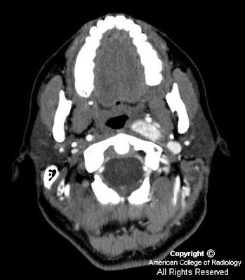
Figure 1 demonstrates a large, intensely-enhancing, heterogeneous left-sided carotid space mass displacing the internal carotid artery (ICA) medially, the external carotid artery (ECA) and its branches antero-medially, and the internal jugular vein (IJV) postero-laterally.
Figure 2 demonstrates anterior displacement of the parapharyngeal fat by the glomus vagale tumor.
Figure 3 demonstrates anterior displacement of the ICA by the left-sided fusiform-shaped glomus vagale tumor.
Figure 4 demonstrates antero-medial displacement of the ICA and posterior displacement of the IJV by the glomus vagale.
Figure 5 with arrows pointing to multiple flow voids within the left carotid space mass.


Diagnosis: Glomus vagale tumor


Paragangliomas account for only 0.6% of all neoplasms of the head and neck. They arise from the paraganlgia or glomus cells, which are part of the extra-adrenal neuroendocrine system, and are named for the paraganglia from which they arise. The four most common sites for paragangliomas of the head and neck are the carotid body (carotid body tumor), the jugular foramen (glomus jugulare), along the path of the vagus nerve (glomus vagale) and in the middle ear (glomus tympanicum). Paragangliomas are characteristically vascular, intensely enhancing lesions that demonstrate low T1 and high T2 signal with multiple flow voids. The "salt-and-pepper" appearance has been named for the T2 appearance, as the high T2 signal of the tumor is the "salt" and the multiple flow voids the "pepper".

Paragangliomas can be distinguished from each other primarily based on their location. The carotid body tumor typically splays the common carotid bifurcation, while the glomus vagale displaces both the ECA and ICA antero-medially away from the IJV. Glomus jugulare tumors characteristically expand and erode the jugular foramen causing the characteristic "moth-eaten" appearance. The glomus tympanicum is a small discrete mass arising from the cochlear promontory in the middle ear.

The current primary treatment modality is surgery with possible pre-operative embolization; radiation is reserved for non-resectable and recurrent cases and those invading the ICA. Paragangliomas have a high (40%-50%) rate of local recurrence, can be locally aggressive with intra-cranial extension (20%), and can behave in a malignant manner (2%-13% metastasize to lung, bones and lymph nodes). Familial paragangliomas (7%-9% of cases) are more commonly multiple (1/3) than sporadic cases (5%), and occur at a younger age. The primary differential diagnosis for a carotid space mass is: nerve sheath tumors, nodal metastasis (renal and thyroid cancer), abscess, and venous thrombosis.

mercoledì 7 dicembre 2005

Joubert syndrome


Findings

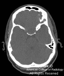
Axial CT shows elongated and thickened superior cerebellar peduncles. Note hte "bat-wing" appearance of the fourth ventricle.
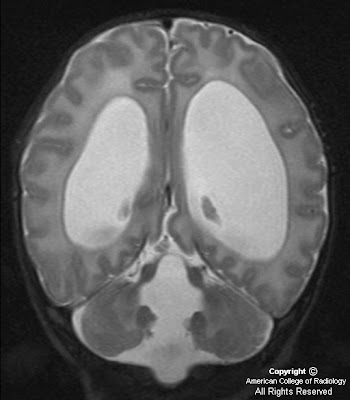
Axial T2-weighted image (Figure 2) through again demonstrates characteristic "bat-wing" appearance of the fourth ventricle with enlarged superior cerebellar peduncles.
Axial T1-weighted inversion recovery image (Figure 3) at the level of the cerebellar peduncles again shows the enlarged superior cerebellar peduncles. The isthmus of the brainstem (the transitional zone between the pons and midbrain) is small and in combination with elongated thickened superior cerebellar peduncles produces the "molar tooth sign." Note the cerebellar hemispheres are in apposition without evidence of fusion.
Mid-sagittal T1-weighted MR (Figure 4) confirms absence of the cerebellar vermis.
Coronal T2-weighted image (Figure 5) shows the thickened cerebellar peduncles (Figure 5) and absence of the normal vermis, without cerebellar hemispheric fusion.


Diagnosis: Joubert syndrome


Joubert syndrome is an autosomal-recessive disorder, characterized by clinical presentation of hypotonia, ataxia, and global developmental delay. A variety of other abnormalities have been described in affected children, primarily episodic hyperpnea, abnormal eye movements, and a characteristic facial appearance. The phenotype may vary even among siblings with Joubert syndrome.

From an imaging perspective, these patients have either complete or partial agenesis of vermis, which results in triangular-shaped mid fourth ventricle and a bat-wing appearance in its superior aspect. The cerebellar hemispheres oppose one another in the midline due to absence of vermis. The isthmus and the midbrain are small in AP diameter, likely secondary to absence of decussation of the superior cerebellar peduncles. This appearance of small brainstem with elongated and thickened superior cerebellar peduncles has been termed the molar tooth sign. It has been proposed that both the superior cerebellar peduncles and the corticospinal tracts remain uncrossed in these patients. The classic imaging findings are, however, not completely specific for Joubert syndrome and have been found recently in a number of very rare congenital syndromes.