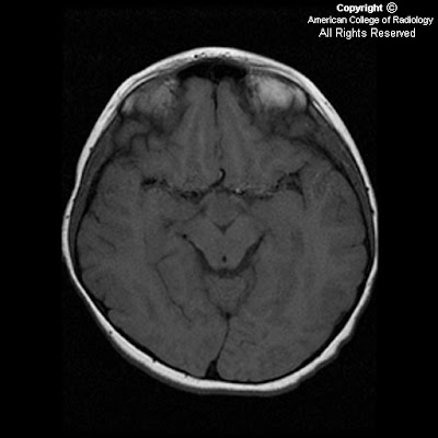


There is an ovoid mass, measuring 1.8 cm anteroposterior x 1.4 cm craniocaudal x 0.8 cm transverse, in the region of the tuber cinerium of the hypothalamus between the infundibular stalk anteriorly and the mamillary body posteriorly (Figure 6). The lesion is seen to extend upwards within the 3rd ventricle (Figures 4 and 5). It is isointense to gray matter signal on T1-weighted images (Figure 1 and Figure 6) and mildly hyperintense on T2 sequences (Figure 2 and 4). No appreciable enhancement is seen on postcontrast administration (Figures 3, 5 and 7).
The pituitary gland appears normal. (Figures 4, 5, 6 and 7).
Diagnosis: Hamartoma of tuber cinerium
Hamartomas of tuber cinerium are nonneoplastic heterotopias of normal neuronal tissue located between the infundibular stalk anteriorly and the mamillary bodies posteriorly. These congenital masses are thought to be due to an anomaly in neural migration between 35 and 40 days of embryonic development, when the hypothalamus is formed.
Hamartomas usually present in young children with two distinct clinical presentations:
- Precocious puberty with symptom onset usually prior to 2 years of age, and
- Seizures - often gelastic type with spasmodic laughter.
- Hamartomas may be sessile or pedunculated. Sessile lesions are almost always symptomatic. These lesions range in size from a few millimeters to 3 to 4 cm. Hamartomas usually demonstrate lack of growth; if growth is detected, surgery or biopsy is indicated. Medical therapy is recommended for sessile lesions with hormonal suppressive therapy (LHRH agonists) and antiseizure medications. Surgery is recommended for pedunculated lesions with refractory symptoms.
On CT, hamartomas appear isodense with brain tissue, with rare calcification and cystic component. MRI demonstrates these lesions to be isointense to gray matter on T1-weighted images and iso- to hyperintense to gray matter on T2-weighted images. The posterior pituitary bright spot is usually preserved. Hamartomas are characteristically nonenhancing; if enhancement is seen, one should consider other diagnoses.
Thin section coronal and sagittal imaging should be performed in any child with precocious puberty or gelastic seizures. The floor of the third ventricle should be smooth from the infundibulum to mamillary bodies. Any nodularity should raise suspicion for a hamartoma in the appropriate clinical setting.
Nessun commento:
Posta un commento