venerdì 25 agosto 2006

Subependymoma


Findings


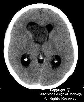
Noncontrast CT images show dilation of the lateral ventricles (Figure 1 and Figure 3) secondary to a large hypodense heterogeneous mass within the right lateral ventricle (Figure 2).
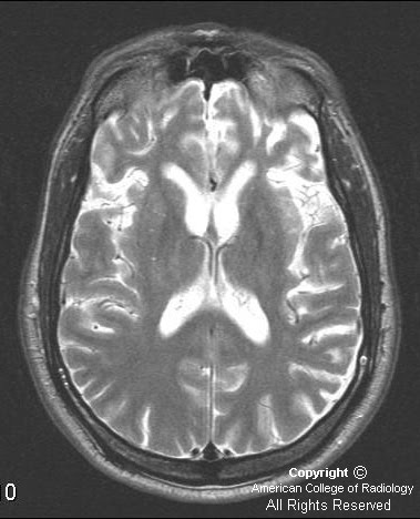
MRI brain with and without contrast. Images show a heterogeneous mass in the right lateral ventricle that causes obstruction of the foramen of Monroe and mild hydrocephalus. The mass is isointense to gray matter on T1 sequences (Figure 6), hyperintense on T2 (Figure 4), shows little if any enhancement on post contrast T1 (Figure 7), and is hyperintense with multiple cystic spaces on FLAIR images (Figure 5).

Differential diagnoses: Intraventricular tumors
- Meningioma
- Choroid plexus papilloma or carcinoma
- Ependymoma
- Subependymoma
- Central neurocytoma
- Metastasis
- Subependymal giant cell astrocytoma


Diagnosis: Subependymoma


Subependymomas are benign intraventricular tumors that arise from cells of the subependymal plate, including subependymal glia, astrocytes and ependymal cells. They are rare, accounting for less than 1% of all intracranial neoplasms. They can occur with ependymomas, which can worsen prognosis. They occur in either the lateral or fourth ventricles, being attached to either septum pellucidum or wall of lateral ventricle, or the ventricular floor, respectively. The larger the lesion, the more likely cystic change and calcification are encountered; however, the calcification is more often punctate and fine rather than coarse, as seen in other intraventricular tumors. Mean age of presentation is 60 years in asymptomatic patients, but only 40 years if symptomatic. There is a male predominance. Surgical resection is often curative, although fourth ventricular subependymomas pose a challenge at total resection.

On CT, an iso- to hypodense intraventricular mass will be seen with cystic change and calcification in larger lesions, and associated hydrocephalus if obstructing. Mild to no enhancement after contrast is typical. On MR, subependymomas are T2/FLAIR hyperintense and T1 isointense with mild to no enhancement with contrast. Because fourth ventricular subependymomas can have calcification and contrast enhancement more typical of ependymomas, diagnostic imaging is more challenging.

Another diagnostic challenge on imaging is the central neurocytoma, a recently described tumor that was routinely mistaken for an intraventricular oligodendroglioma on histologic analysis. These intraventricular tumors have a striking resemblance to a subependymoma on CT and MR images. Neurocytomas, however, more reliably enhance after contrast administration, and have a more heterogeneous cystic appearance. They also are usually found in younger patients.

mercoledì 23 agosto 2006

Pick's disease


Findings

Figure 1, Figure 2, Figure 3, and Figure 4: MR images demonstrate marked atrophy involving the frontal and temporal lobes on T1, T2, and FLAIR.
Figure 5: Selected axial SPECT images after the intravenous administration of Tc-99m Neurolite demonstrate decreased uptake in the frontal and temporal lobes, right slightly greater that left.


Diagnosis: Pick's disease


Pick's disease is described as a rare, progressive, degenerative disease, mostly involving the frontal and temporal lobes. This was first described by Arnold Pick in 1892. It is not thought to be inherited. Women are more affected than men. There is no cure.

This disease is more common in a younger age group (40 to 60) with an average of 54. Alzheimer's disease would be more likely in older patients, over 70.

Behavioral and physical changes include repetitive or compulsive behaviors, mood changes, apathy, echolalia, aphasia, apraxia, rigidity, and a multitude of other behaviors that range from not speaking or moving to rapid pacing. It will always progress to a vegetative state.

MRI will demonstrate atrophy of the frontal and temporal lobes with preservation of the pre- and postcentral gyrus on all sequences. There is no abnormal enhancement associated with this disease.

SPECT imaging will demonstrate decreased radiotracer uptake in the frontal and temporal lobes.

Histologic evaluation, which is usually postmortem, demonstrates a "ballooned" appearance of the neurons related to swelling. These are called Pick bodies. This is different than Alzheimer's type dementia, which demonstrates amyloid plaques and neurofibrillary tangles. In multi-infarct dementia, there is myelin and axonal loss with associated astrocytosis and generalized necrosis.

venerdì 18 agosto 2006

Hamartoma of tuber cinerium


There is an ovoid mass, measuring 1.8 cm anteroposterior x 1.4 cm craniocaudal x 0.8 cm transverse, in the region of the tuber cinerium of the hypothalamus between the infundibular stalk anteriorly and the mamillary body posteriorly (Figure 6). The lesion is seen to extend upwards within the 3rd ventricle (Figures 4 and 5). It is isointense to gray matter signal on T1-weighted images (Figure 1 and Figure 6) and mildly hyperintense on T2 sequences (Figure 2 and 4). No appreciable enhancement is seen on postcontrast administration (Figures 3, 5 and 7).
The pituitary gland appears normal. (Figures 4, 5, 6 and 7).


Diagnosis: Hamartoma of tuber cinerium


Hamartomas of tuber cinerium are nonneoplastic heterotopias of normal neuronal tissue located between the infundibular stalk anteriorly and the mamillary bodies posteriorly. These congenital masses are thought to be due to an anomaly in neural migration between 35 and 40 days of embryonic development, when the hypothalamus is formed.

Hamartomas usually present in young children with two distinct clinical presentations:
- Precocious puberty with symptom onset usually prior to 2 years of age, and
- Seizures - often gelastic type with spasmodic laughter.
- Hamartomas may be sessile or pedunculated. Sessile lesions are almost always symptomatic. These lesions range in size from a few millimeters to 3 to 4 cm. Hamartomas usually demonstrate lack of growth; if growth is detected, surgery or biopsy is indicated. Medical therapy is recommended for sessile lesions with hormonal suppressive therapy (LHRH agonists) and antiseizure medications. Surgery is recommended for pedunculated lesions with refractory symptoms.

On CT, hamartomas appear isodense with brain tissue, with rare calcification and cystic component. MRI demonstrates these lesions to be isointense to gray matter on T1-weighted images and iso- to hyperintense to gray matter on T2-weighted images. The posterior pituitary bright spot is usually preserved. Hamartomas are characteristically nonenhancing; if enhancement is seen, one should consider other diagnoses.

Thin section coronal and sagittal imaging should be performed in any child with precocious puberty or gelastic seizures. The floor of the third ventricle should be smooth from the infundibulum to mamillary bodies. Any nodularity should raise suspicion for a hamartoma in the appropriate clinical setting.

giovedì 17 agosto 2006

Lemierre syndrome


Findings

Figure 1: The CT scan of the neck demonstrates low density material within the right internal jugular vein. This is consistent with internal jugular vein thrombosis.
The CT scan of the chest demonstrates numerous peripheral nodules, many with central cavitation, as well as bilateral pleural effusions and a left lower lung consolidation. The presence of multiple peripheral nodules, some of which are cavitary, as well as consolidation and pleural effusions, is very consistent with septic embolic disease. In this patient, the septic emboli are arising from a thrombophlebitis of the internal jugular vein.


Diagnosis: Lemierre syndrome


Lemierre syndrome, also known as postanginal sepsis, is thrombophlebitis of the internal jugular vein with metastatic septic emboli, generally following an acute oropharyngeal infection.

The disease typically progresses in a stepwise fashion, starting with a primary infection, which is usually a pharyngitis of the palatine tonsils and peritonsillar tissue (in almost 90% of cases). The infection then invades the parapharyngeal space, usually via lymphatic vessels. The posterior parapharyngeal space contains the internal jugular vein, common carotid artery, cervical sympathetic trunk, and cranial nerves IX to XII. Although the infection can involve any of these structures, the most common consequence is internal jugular vein thrombophlebitis. Clinically, patients may have pain or swelling of the neck, sometimes with associated trismus or spasm of the sternocleidomastoid muscle. Once the internal jugular vein is involved, the infection can spread hematogenously to any part of the body, but most frequently affects the lungs, with metastatic infection occurring in 80% of cases, and to the joints, particularly the hips, shoulders, and knees, in 16% of cases. Although findings of pulmonary septic emboli, including multiple peripheral cavitating nodules or pulmonary infarction, are the classic presentation of Lemierre syndrome, less specific pulmonary manifestations, such as noncavitating pulmonary infiltrates and pleural effusions, are frequently seen as well.

Treatment involves a prolonged course of high-dose intravenous antibiotics. Prior to the use of antibiotics, this disease carried a mortality rate of 90%. Although the incidence of this disease has decreased substantially, delay in treatment is associated with significant morbidity and mortality. Therefore, familiarity with this syndrome is important so the diagnosis can be made quickly and appropriate antibiotic coverage started. The constellation of internal jugular vein thrombosis with pulmonary findings, even if they are not clearly septic emboli, should immediately lead to the consideration of Lemierre syndrome as the diagnosis until proven otherwise. Although a clinical history of recent pharyngitis, or evidence of an inflamed pharynx is helpful, as Hall warned in 1939, “Be not deceived by a comparatively innocent-appearing pharynx, as the veins of the pharynx may be carrying the death sentence for your patient.”

lunedì 14 agosto 2006

Enlarged vestibular aqueduct


Findings

Figure 1: Axial CT demonstrates enlarged vestibular aqueducts bilaterally.
Figure 2: Axial CT shows the cochlea and normal ossicles.


Diagnosis: Enlarged vestibular aqueduct syndrome


An enlarged vestibular aqueduct is defined by an anterior-posterior diameter greater than 1.5 mm, at the midpoint between its external aperture and the common crus. Hearing loss usually is bilateral, progressive in nature, and may be associated with vertigo. Some cases may be familial.

This is thought to occur as a result of arrest of inner ear development during the fifth week of embyronic life. Clinically, these patients hear normally during the first few years of life, with progressive sensorineural hearing loss developing as late as the teenage years. Vestibular aqueduct syndrome may be unilateral or bilateral.

It is suggested that the progression of hearing loss is often triggered by head trauma and activities that cause sudden fluctuation in cerebrospinal fluid pressure. The large vestibular aqueduct would allow back pressure to the inner ear, resulting in damage to the membranous labyrinth.

On MRI, the content of the large vestibular aqueduct becomes visible: dilatation of the endolymphatic duct and sac is seen, although their size is not always proportional to the dilatation of the bony vestibular aqueduct.

Large vestibular aqueduct syndrome is seen in Pendred’s syndrome, an autosomal recessive condition characterized by deafness and goiter. In comparison, a narrowed vestibular aqueduct less than 0.5 mm in diameter may be seen with Meniere disease.


Axial CT shows an example of a normal caliber vestibular aqueduct.

giovedì 3 agosto 2006

Spinal epidural capillary hemangioma


Findings

On axial and sagittal T1-weighted images (Figure 1 and Figure 4), the lesion is slightly hypointense compared with the spinal cord. There is marked compression of the spinal cord. On the sagittal T2-weighted image (Figure 2), the lesion is hyperintense relative to the spinal cord. After the administration of gadolinium (Figure 3 and Figure 5), the lesion enhances intensely and homogeneously, which is typical for a hemangioma.


Diagnosis: Spinal epidural capillary hemangioma


Purely epidural hemangiomas account for up to 12% of all spinal hemangiomas, the large majority of which are located in the vertebral bodies, with occasional epidural extension. When purely epidural hemangiomas occur, they tend to develop in the thoracic spine, less often in the lumbosacral spine, and only rarely in the cervical spine. Extension to the paravertebral space with neural foraminal widening and adjacent bony erosion is common. The lesion is generally located in the posterior spinal canal, and frequently partially encircles the spinal cord. They are usually solitary lesions, but may be associated with hemangiomas in other parts of the body, including cutaneous vascular lesions.

Symptoms tend to be gradual in onset, and include back pain, leg weakness, ataxia, parasthesias, and eventually paraparesis. Symptoms may be brought on acutely, most often secondary to trauma. Microhemorrhage in the lesion, which may occur with even minor trauma, can cause an increase in mass effect of the lesion with sudden worsening of, or acute onset of symptoms. Exercise, systemic infection, pregnancy, rapid change in posture, or straining can also cause acute onset of symptoms.

The differential diagnosis for capillary or cavernous epidural hemangioma includes lymphoma, meningioma, neurogenic tumors, epidural hematoma, epidural abscess, metastatic disease, and extramedullary hematopoiesis. Epidural hemangiomas tend to have very consistent imaging characteristics, and are generally isointense to the spinal cord on T1-weighted images, hyperintense on T2-weighted images, and demonstrate intense, homogeneous enhancement after administration of contrast. This specific constellation of imaging characteristics may help distinguish an epidural hemangioma from the other lesions in the differential diagnosis. In addition, a thin rim of low-signal intensity is often visible surrounding the lesion on both T1- and T2-weighted images, which often helps establish the lesion to be truly epidural, rather than extramedullary but intradural.

Treatment involves surgical resection, and complete resection often results in complete neurologic recovery. However, intraoperative bleeding (due to the vascular nature of the tumor) or intrathoracic extension may prevent complete excision.

mercoledì 2 agosto 2006

Bilateral persistent stapedial artery (PSA)


Findings

Figure 1 and Figure 2: Axial images of temporal bone show presence of bilateral persistent stapedial artery (PSA) coursing over the cochlear promontory.
Coronal image of temporal bone shows the origin of the PSA from the petrous part of the carotid artery and coursing superiorly along the cochlear promontory. Tympanic portion of the facial nerve is also seen.


Diagnosis: Bilateral persistent stapedial artery (PSA)


PSA is a rare congenital vascular anomaly of the middle-ear. It is usually asymptomatic and an incidental finding at surgery, or presents with pulsatile tinnitus or conductive hearing loss. The PSA arises from the petrous part of the internal carotid artery, enters the anteromedial hypotympanum, and is contained in the Jacobson canal. After leaving the osseous canal, it crosses the cochlear promontory and passes through the obturator foramen of the stapes. It then enters the fallopian canal through a dehiscence just behind the cochleariform process and travels anteriorly in the anterior facial canal. This leads to prominence of the tympanic part of the facial nerve; an indirect imaging sign of PSA. Finally, the PSA exits just before the geniculate ganglion, entering into the extradural space of the middle-cranial fossa. Another indirect sign of PSA is absent or hypoplastic foramen spinosum, though the foramen spinosum can be congenitally absent in 3.2 % of the population; hence, other imaging signs of PSA need to be present to confirm its presence.

A PSA can be associated with an aberrant internal carotid artery or other middle-ear anomalies, especially of the stapes and facial nerve. There have been very few rare cases reported of bilateral PSA (1, 2), including one reported case of bilateral PSA with bilateral aberrant carotid arteries (3).