mercoledì 31 gennaio 2007
Arachnoid cyst
Findings
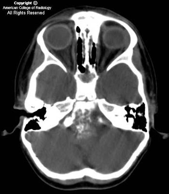
There is a small left frontal extra-axial hypodense fluid collection with associated thinning of the calvarium. No significant mass effect on the brain parenchyma. No acute intracranial abnormalities.
Differential diagnosis:
- Arachnoid cyst
- Chronic subdural hematoma
- Subdural hygroma
- Epidermoid cyst
- Prominent CSF space
- Porencephalic cyst
- Neuroglial cyst
Diagnosis: Arachnoid cyst
Key points
An arachnoid cyst is a benign, intra-arachnoid, CSF-filled cyst that does not communicate with the ventricular system. Arachnoid cysts are fairly common, accounting for approximately 1% of all intracranial masses. They are often asymptomatic and are found incidentally, but are twice as common in imaging performed for history of seizure. If present, symptoms vary with the size and location of the cyst and can include, headache, sensorineural hearing loss, dizziness, and even obstructive hydrocephalus. Arachnoids cysts are usually stable in size but have been known to slowly enlarge. No treatment is usually required, but resection, fenestration, or shunt placement may be performed for serious symptoms.
Radiology
In general: Arachnoid cysts may appear anywhere in the cranium, although the middle cranial fossa is the most common location. They can vary in size from only a few millimeters to ten centimeters or more. They typically displace cortex and may even "buckle" the gray-white border.
CT: CSF density, usually associated thinning/remodeling of adjacent bone, no enhancement with contrast
MR:
- T1WI - isointense with CSF, sharp margins
- T2WI – isointense with CSF
- FLAIR – suppresses completely
- DWI – no restricted diffusion
- T1+C – no enhancement
lunedì 29 gennaio 2007
Vein of Galen aneurysm
Findings
There is a large vascular structure just posterior to the third ventricle and superior to the cerebellar vermis. There is some mass-effect on the third ventricle with dilatation of the third ventricle and frontal horns. No definite intracranial hemorrhage noted. MRI shows a large tubular structure with flow identified in the region of the vein of Galen extending into the straight sinus towards the torcula Herophili. There are numerous collateral vessels within the ambient and perimesencephalic cisterns as well as surrounding the brainstem.
Diagnosis: Vein of Galen aneurysm
Key points
Vein of Galen "aneurysm" is actually an AVM with an associated varix of the vein of Galen.
Two main types exist
- Vein of Galen AVM.
- Vein of Galen aneurysmal dilatation.
Vein of Galen AVM is the result of a developmental malformation that involves the vein of Galen. It is an A-V shunt in the wall of an embryologic venous precursor (Median Vein of the Prosencephalon [MVP]) and in this situation a normal vein of Galen does not exist and is replaced by a persistent MVP. The MVP does not drain normal brain tissue. In other words, a Vein of Galen AVM is not anatomically associated with a parenchymal AVM. The dilated MVP drains via the dural sinuses. Vein of Galen AVM presents in infancy with high output cardiac failure and macrocephaly. The A-V fistulas may be single or multiple. Intrauterine diagnosis is usually possible by doppler. Intracranial hemorrhage is rare.
Vein of Galen AVM should be differentiated from aneurysmal dilatation of the vein of Galen which occurs in association with a parenchymal AVM or the rare asymptomatic varicose vein of Galen.
Therapy: Endovascular embolization via an arterial or venous access.
Radiology
Transcranial doppler sonogram shows an anechoic structure in the superoposterior aspect of the third ventricle with flow.
CT, MRI and MRA show the vascular abnormality and assess the brain parenchyma for abnormalities.
Conventional angiography is essential to assess the malformation and to plan the therapy.
Etichette:
AuntMinnie,
Malformations,
Neuro,
Vascular
giovedì 25 gennaio 2007
Nevoid Basal Cell Carcinoma (Gorlin) Syndrome (NBCCS)
Findings
Figure 1: Characteristic calcification of the petroclinoid ligaments and the diaphragma sellae is present.
Figure 2: In addition, there are dural calcifications involving the falx cerebri and tentorium cerebelli.
Figure 3: Soft tissue lesion involving the right scalp is also visualized and corresponds to pathologically proven basal cell carcinoma.
Figure 4: There is subtle parallel orientation of the bodies of the lateral ventricles that suggests dysgenesis of the corpus callosum, a finding that may occur in as many as 10% of patients with NBCCS.
Diagnosis: Nevoid Basal Cell Carcinoma (Gorlin) Syndrome (NBCCS)
NBCCS (Gorlin syndrome) is a rare autosomal dominant disorder characterized by multiple basal cell carcinomas, odontogenic keratocysts of the jaw, palmar/plantar pits, calcification of the falx cerebri, and rib and spine anomalies. Patients with NBCCS have a propensity to develop multiple neoplasms such as basal cell carcinoma (>90%), medulloblastoma (10%), meningioma, and ovarian or cardiac fibromas.
Radiologic evaluation is important in patients suspected of having NBCCS because the identification of a pathogenic mutation is not always possible, and the clinical manifestations may be subtle, especially in children or African Americans. Recognition is also clinically important as these patients are sensitive to ionizing radiation and have been known to develop iatrogenic malignancy as a result of radiation therapy.
Key radiologic features include:
- Extensive calcification of the falx cerebri, tentorium cerebellum, diaphragm sella, or petroclinoid ligaments
- Odontogenic keratocyst of the jaw
- Bifid, splayed, fused, or absent ribs, especially involving the 3rd to 5th ribs
- Scoliosis, hemivertebrae, or fused vertebrae
- Flame-shaped lucencies of the hands or feet
- Scapular abnormalities
Nevoid Basal Cell Carcinoma Syndrome includes two Major critera or one Major and two Minor criteria
Major Criteria
- More than two basal cell carcinomas or one basal cell carcinoma under the age of 20
- Odontogenic keratocysts of the jaw
- Three or more palmar or plantar pits
- Lamellar calcification of the falx cerebri
- Rib Anomalies (bifid, synostosed, hypoplastic)
- First degree relative with NBCCS
Minor Criteria
- Spina bifida occulta or other vertebral anomalies
- Brachymetacarpaly in at least one limb
- Hypertelorism or telcanthus
- Frontal bossing
- Sprengel deformity
- Ovarian fibroma
- Medulloblastoma
- Meningioma
- Flame shaped lucencies of the phalanges
- Bridging of the sella turcica
- Mesenteric cysts
- Cardiac fibroma
- Dysgenesis/agenesis of the corpus callosum
Etichette:
ACR,
Genetic-Metabolic,
Neoplasm,
Neuro,
Systemic
venerdì 19 gennaio 2007
Third Ventricle Arachnoid Cyst
Findings
Figure 1 and Figure 2: Axial and coronal T2 show a large cystic lesion, located within the third ventricle. There is mass effect on the surrounding normal brain structures.
Figure 3 and Figure 4: Postcontrast axial and sagittal T1 show no postcontrast enhancement of the lesion.
Figure 5: The lesion is dark (negative) on the DWI sequence.
Diagnosis: Third Ventricle Arachnoid Cyst
Arachnoid cysts form when the two layers of the arachnoid membrane split and a potential cavity is formed. The cavity fills with cerebrospinal fluid by an unknown mechanism, thought to be CSF pulsations. They can continue to grow as CSF flows in and is trapped. Arachnoid cysts rarely hemorrhage in trauma cases.
Arachnoid cysts typically cause symptoms secondary to mass effect on surrounding structures. Alternatively, they may remain asymptomatic and be discovered only as an incidental finding. Obstructive hydrocephalus, with increased intracranial pressure resulting in headaches, seizures, and developmental delay, is a common presenting feature.
Imaging findings typically feature a cystic structure with signal characteristics of CSF and dark signal on diffusion-weighted imaging; rarely, some evidence of hemorrhage may be present, especially in the setting of trauma. No solid component is present, and there is no postcontrast enhancement. There may be associated calvarial inner table scalloping, and there is typically mass effect on surrounding normal structures. Features that distinguish this entity from a subdural hygroma are convex borders, bony remodeling, and lack of substantial hemorrhage. Both entities typically cause sulcal effacement and mass effect. In cases of underlying atrophy/encephalomalacia, there would be no bone remodeling (exception is a porencephalic cyst), no surrounding normal brain architecture, and sulcal enlargement rather than effacement; cerebral veins would be seen coursing through the CSF, rather than being displaced (as with a subdural hygroma or arachnoid cyst). The lack of positive signal on the diffusion-weighted images and absence of a solid component, especially of fat signal, distinguishes an arachnoid cyst from an epidermoid, which would typically be bright on DWI.
mercoledì 17 gennaio 2007
Warthin's tumor
Findings
Figure 1: Contrast enhanced axial CT scan image showing the predominantly cystic left intraparotid mass, with enhancing eccentric nodule and minimal wall thickening. Right parotid gland is normal. No cervical lymphadenopathy was seen on the study.
Differential diagnosis:
- Pleomorphic adenoma
- Warthin’s tumor
- Lymphoma
- Lymphoepithelial cyst
- Mucoepidermoid carcinoma
- Adenoid cystic carcinoma
- Metastasis
Diagnosis: Warthin's tumor
Also known as papillary cystadenoma lymphomatosum, Warthin’s tumors exhibit a slight male predominence and most commonly occur in patients older than 50. The majority of cases are seen in patients in their seventh to eighth decade of life. They are the second most common benign parotid tumor next to pleomorphic adenoma (approximately 75% to 80% of benign parotid tumors are pleomorphic adenoma). Smokers have an eightfold increased risk of developing this tumor, which likely accounts for the historic male predominance. Warthin's tumors are the most common bilateral parotid tumors. About 6% to 10% of patients will have bilateral tumors. They most commonly arise in the tail of the parotid gland, usually presenting as painless and slow-growing masses. There is a very low incidence of malignant transformation.
Typical CT characteristics include a small, ovoid, well-circumscribed, smoothly marginated mass of the posterior parotid gland. The tumor can be cystic or lymphoma-like in appearance. Warthin’s tumors often present as homogeneous soft tissue densities. Cyst formation is common, and the classic appearance is of a cystic lesion with focal tumor nodule(s). On T1-weighted MR, the mass contrasts to the hyperintense signal of the normal parotid gland. On T2WI, Warthin’s tumors are often heterogeneous, with variable signal intensity. Warthin’s tumors show significant uptake of Technetium 99m pertechnetate, as does oncocytoma.
Warthin’s tumor is usually treated by surgical excision. Facial nerve involvement is rare and the facial nerve can usually be spared. Conservative management with close follow-up is acceptable, particularly in poor surgical candidates.
giovedì 11 gennaio 2007
Trigeminal schwannoma
Findings
CT postcontrast reveals an unenhancing soft tissue mass centered at Meckel's cave on the left (Figure 1)
CT bone window (Figure 2) reveals a markedly enlarged foramen ovale on the left, with smooth, corticated margins.
Axial T1-WI (Figure 3) demonstrates a well-circumscribed extraaxial mass in the region of left Meckel’s cave that is mildly hypointense to brain parenchyma. The mass shows heterogeneous signal intensity on FLAIR (Figure 3). Postcontrast images show moderately intense enhancement, extending into the cisternal segment of cranial nerve V (Figure 4). The mass is seen to extend through a widened foramen ovale into left infratemporal fossa and present as masticator mass (Figure 4).
Diagnosis: Trigeminal schwannoma
Schwannomas account for 6% to 8% of intracranial neoplasms. Vestibular schwannomas are the most common cranial nerve schwannomas, followed by trigeminal and facial schwannomas.
The trigeminal nerve (CN V) exits the brainstem at the level of the mid pons, and its three divisions—the ophthalmic (CN V1), maxillary (CN V2), and mandibular (CN V3) branches—together proceed anteriorly toward the trigeminal ganglion in the Meckel’s cave. From here, the mandibular division exits inferiorly via the foramen ovale. The maxillary and ophthalmic divisions continue anteriorly along the lateral aspect of the cavernous sinus. Eventually, the ophthalmic division enters the orbit via the superior orbital fissure, while the maxillary division exits the cranial vault through the foramen rotundum.
Trigeminal schwannomas are encapsulated tumors of Schwann cells displace rather than infiltrate fascicles of CN V3.
Trigeminal schwannomas usually present in the third to fourth decade with sensory disturbances. Mastication muscles weakness or facial pain is less common.
Trigeminal schwannomas may arise along any segment of CN V, but the majority develop at the level of trigeminal ganglion. They may then extend posteriorly into the posterior fossa, or anteriorly through the skull base foramina, and often have a “dumbbell” configuration.
On nonenhanced CT scans, most schwannomas are isodense relative to brain parenchyma. Calcification or areas of hemorrhage are rare. On contrast-enhanced CT scans, the enhancement pattern is typically homogeneous. Foramen ovale is most commonly enlarged by CN V3 schwannoma.
Schwannomas typically are isointense or slightly hypointense relative to gray matter on T1-weighted images and hyperintense on T2-weighted images. Gadolinium enhancement typically is homogeneous, although larger schwannomas can show areas of cystic degeneration and heterogeneous signal intensity. Atrophy of masticator muscles may be seen as high signal fatty infiltration on T1-weighted images with volume loss.
Surgical resection is the treatment of choice.
Trigeminal schwannoma may be associated with Neurofibromatosis type 2, especially if it occurs in younger patients.
venerdì 5 gennaio 2007
Supratentorial Primitive Neuroectodermal Tumor (S-PNET)
Findings
There is a mass in the left anterior parietal lobe measuring 3.2 x 3.0 cm in maximum transverse dimensions. The mass is heterogeneous and demonstrates a central area of increased T1 signal. There is minimal surrounding edema. Mild peripheral heterogeneous enhancement is evident on the T1-weighted sequence.
There is a large mass in the left parietal lobe on the Axial T1 FLAIR with contrast image (Figure 1) that demonstrates heterogenous enhancement. The red arrow demonstrates the left central sulcus.
There is a large, complex, hemispheric mass with minimal peritumoral edema on the Axial T2 FRFSE image (Figure 3).
The green arrows on the axial diffusion-weighted image (Figure 4) demonstrate areas of increased signal (resticted diffusion) centrally within the mass.
Diagnosis: Supratentorial Primitive Neuroectodermal Tumor (S-PNET)
Primitive neuroectodermal tumors (PNETs) are differentiated by the anatomical location. The most common location for a PNET is in the posterior fossa and is called a medulloblastoma (MB). PNETs found anywhere else in the brain are considered supratentorial primitive neuroectodermal tumors (S-PNETs). The histological appearance of medulloblastomas and S-PNETs are identical, with medulloblastomas having a better prognosis than S-PNET. (S-PNET > 30-35% 5 year survival; medulloblastoma > 80-85% 5-year survival). Critical survival factors include absence of metastases, patient age > 2 years, and smaller, solid tumors as necrosis suggest a worse prognosis.
Supratentorial primitive neuroectodermal tumors (S-PNETs) are rare tumors accounting for less than 1% of all primary CNS tumors and are more commonly found in young children in the first five years of life. According to the WHO classification, S-PNET is defined as an embyronal tumor in the cerebrum or suprasellar region consisting of undifferentiated or poorly differentiated neuroepithelial cells that display or have the potential for divergent differentiation.
S-PNETs do not have a specific clinical presentation but vary with the size and location of the tumor. Clinical presentation varies from seizures, to motor deficits, to visual disturbances. Paresis and seizures can occur when the tumor involves the cortex. Headaches, nausea, and vomiting are symptoms of increased intracranial pressure seen when the flow of CSF is obstructed. Endocrinopathies or visual deficits may result from suprasellar tumors. S-PNETs can be found in the pineal region, where they cause ventricular obstruction and gaze/convergence difficulties; in the cerebral hemispheres, where they are usually larger at the time of diagnosis with a mean diameter of approximately 5 cm; or in the suprasellar region, where they tend to be smaller due to neuroendocrine and visual disturbances.
giovedì 4 gennaio 2007
Occipital encephalocele
Findings
There is a large occipital encephalocele, containing mixed solid and cystic areas. The calvarial defect is between 1-2 cm, through which brain tissue is seen to herniate
Differential diagnosis:
- Occipital encephalocele
- Cystic hygroma
- Teratoma
- Branchial cleft cyst
- Scalp edema
- Epidermal scalp cyst
Diagnosis: Occipital encephalocele
Key points
An encephalocele results from failure of the surface ectoderm to separate from the neuroectoderm. The final result is a bony defect in the skull table, which allows herniation of the meninges (cranial meningocele) or herniation of brain tissue.
Encephaloceles represent 10-15% of all neural tube defects.
Of encephaloceles, occipital encephaloceles are most common (75-80%). 90% are midline.
The absence of brain tissue within the herniated sac is the single most favorable prognostic feature for survival.
Incidence ranges from 1 in 3000 (Southeast Asia) to 1 in 10,000 live births (North America).
Once an encephalocele is diagnosed, a search for associated intracranial and extracranial abnormalities (60-80%) should be performed. 15-20% have additional severe CNS defects including additional neural tube defects, Dandy-Walker malformation, Chiari malformation, hydrocephalus and optic nerve malformations.
The risk of chromosomal abnormalities is 13-44%; therefore, karyotyping should be offered to the mother. Most common chromosomal anomaly is Trisomy 18.
Associated with genetic syndromes such as Meckel-Gruber, von Voss, Chemke, Roberts, and Knobloch syndromes. Important to evaluate digits for polydactyly, kidneys, and for oligohydramnios to exclude Meckel-Gruber syndrome.
Diagnosis is thought to be impossible before skull ossification starts at 10 weeks gestation. Earliest U/S diagnosis has been at 13 weeks.
Etichette:
AuntMinnie,
Malformations,
Neuro,
Pediatric
martedì 2 gennaio 2007
Chordoma
Findings
CT (Figure 1 and Figure 2) shows an expansile, destructive mass originating from the clivus. There is destruction of the anterior arch of C1-vertebra, and the mass extends through the foramen magnum (Figure 3 and Figure 4). Mass effect with compression is seen on the pons and cerebellum. High-attenuation foci are noted within the tumor mass. The bulk of the tumor is hyperdense relative to the adjacent neural axis.
The lesion is iso- to hypo-intense on T1-weighted image (Figure 5) and iso-intense on T2-weighted image (Figure 6). Moderate heterogeneous enhancement is seen within the lesion (Figure 7 and Figure 8). The pituitary gland is normally visualized.
Diagnosis: Chordoma
Chordomas are rare, slow-growing malignant neoplasms that originate from embryonic remnants of the primitive notochord. After birth, the only remaining normal notochordal elements are the nuclei pulposi. However, notochordal remnants may remain along the neural axis.
The common locations of chordomas are:
- sacrococcygeal (50%)
- clival (35%)
- vertebral body (15%)
The peak incidence of chordomas is in the fifth to sixth decade. These are rare in children and usually arise in the spheno-occipital region.
Metastatic spread of chordomas is observed in 7%-14% of patients with lymph node, pulmonary, bone, cerebral, or abdominal visceral involvement, predominantly from massive tumors.
Chordomas appear as expansile, multilobulated, well-circumscribed, destructive midline masses. High attenuation foci are noted within the tumor matrix in 50 % representing ossific fragments. Heterogeneous enhancement is seen with areas of cystic necrosis mixed with enhancing soft tissue. These tumors classically show high-signal intensity on T2-weighted images. Vascular encasement and displacement may be seen in 80% of cases.
Complete surgical resection is the treatment of choice and often combined with radiation therapy. Local recurrence is common, despite combined therapy.
Etichette:
ACR,
Head - Neck,
Neoplasm,
Neuro,
Pediatric
Iscriviti a:
Post (Atom)