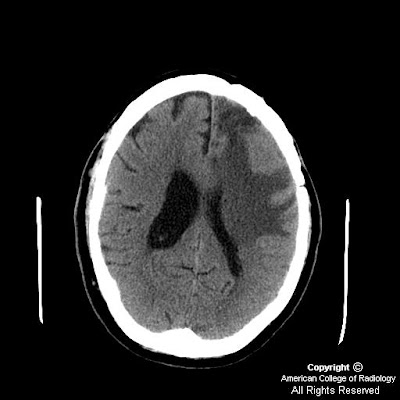


Findings
CT: Post-craniotomy surgical changes with vasogenic edema extending from left frontal lobe into left parietal and temporal lobes. There is symmetric cerebellar atrophy without evidence of asymmetric right hemispheric volume loss.
PET/CT: Decreased metabolism in left frontal lobe and right cerebellar hemisphere representing crossed cerebellar diaschisis.
MR: Enhancement throughout much of the remaining left frontal lobe, with extension into the right frontal lobe, basal ganglia, and the genu of the corpus callosum.
MRI: While PET/CT and MRI findings were suggestive of radiation necrosis, tissue biopsy showed evidence of recurrent malignant oligoastrocytoma (WHO grade 3). Cerebral lesion was subsequently resected surgically.
Diagnosis: Crossed Cerebellar Diaschisis (CCD) in a patient with radiation necrosis and recurrent malignant oligoastrocytoma (WHO grade 3)
Crossed cerebellar diaschisis (CCD) is a functional reduction in the cerebellar hemisphere contralateral to a cerebral lesion. It has been observed following strokes, intracranial tumors, epilepsy, and various types of intracranial trauma. There is a direct relationship between the incidence of CCD and increasing lesion size. CCD can range from a potentially reversible finding (typically following an acute stroke) to permanent degeneration. While most frequently associated with frontal lobe pathology, CCD can also be present with lesions involving the thalamus, basal ganglia, and the internal capsule. The cerebropontocerebellar pathway is thought to be the etiology of CCD; its interruption leading to a reduction in oxygen metabolism and glucose uptake in the contralateral cerebellar hemisphere that can be demonstrated with PET imaging. Conventional CT and MRI studies often do not show any abnormalities, as diaschisis does not always produce structural alterations.
Nessun commento:
Posta un commento