venerdì 29 ottobre 2010

Pott's Puffy Tumor


Findings

Figure 1: The image shows a large collection anterior to the frontal bone and a large epidural abscess with peripheral enhancement.
Figure 2: Erosion of the frontal bone. Sinus tract of the bone into the large anterior collection.
Figure 3: Large collection anterior to the frontal bone.
Figure 4: Large epidural abscess.
Figure 5: Large epidural abscess with peripheral enhancement.
Figure 6: Large collection anterior to the frontal bone. Large epidural abscess. Normal bone marrow signal. Abnormal bone marrow signal in the frontal bone.
Figure 7: Large collection anterior to the frontal bone with peripheral enhancement. Large epidural abscess with peripheral enhancement.
Figure 8: Abnormal bone marrow signal in the frontal bone. There is a lack of enhancement of the frontal bone. The abnormal dark bone marrow signal on T1 images and lack of enhancement on T1 post gad fat sat is consistent with dead necrotic bone from severe osteomyelitis.


Diagnosis: Pott's Puffy Tumor


Pott’s puffy tumor is a subperiosteal abscess of the frontal bone that appears as a localized swelling of the forehead associated with frontal osteomyelitis. Pott’s puffy tumor is a complication of frontal sinusitis or trauma, which is predominatly seen in the adolescent age group. However, there are a few case reports in adults. Pott’s puffy tumor is a rare complication of frontal sinusitis in the post antibiotic era but can be seen in patients with undiagnosed or partially treated sinusitis. Patients will typically present with frontal scalp swelling, headache, fever, nasal drainage, and frontal sinus tenderness. Ocassionally, Pott’s puffy tumor can mimic findings of preseptal or orbital cellulitis. In severe cases, there will be neurologic decompensation. Varying degrees of hemiparesis, obtundation, papillary dilatation or aphasia have been described in case reports.

Imaging is necessary to exclude intracranial complications such as epidural abscess. Pott’s puffy tumor can also be associated with dural sinus thrombosis, meningitis, subdural empyema, epidural abscess, brain abscess, and rarely seizure. Intracranial infection is caused by posterior extension from the frontal sinus while preseptal and orbital cellulitis is caused by downward spread from the frontal sinus to the orbit. Younger children who do not have pneumatized frontal sinuses, are more likely to have ethmoid sinusitis. Orbital cellulitis is a more common complication in patients with ethmoid sinusitis.

Patients must be treated with a combination of surgery and long-term antibiotic therapy.

lunedì 25 ottobre 2010

Infected 4th Branchial apparatus cyst


Findings

Figure 1: Neck CT, contrast enhanced, at level of pyriform sinus. The left pyriform sinus is effaced by an inflammatory mass.
Figure 2: Neck CT, contrast enhanced, at level of subglottic trachea. The image demonstrates continuation of the large inflammatory mass with small areas of necrosis or abscesses. Note displacement of the trachea to the right and lateral displacement of the carotid sheath vessels. Reactive lymphadenopathy is present in the internal jugular chain.
Figure 3: Neck CT, contrast enhanced, at level of the thyroid gland. The image shows a mixed attenuation mass in the enlarged left lobe of the thyroid. This mass arises from extention of the extrinsic anterior and lateral inflammatory mass with phlegmon and abscesses from an infected 4th branchial apparatus cyst.
Figure 4: Neck CT coronal reformation, contrast enhanced. There is extensive phlegmon with multiloculated abscesses, extending from the left lower pharyngeal wall into the left lobe of the thyroid gland. Reactive lymphadenopathy in the left internal jugular lymph node chain is present.


Diagnosis: Infected 4th Branchial apparatus cyst


The main differential diagnostic considerations for a cystic neck mass in children include suppurative lymph nodes, abscess, thyroglossal duct cyst, lymphatic malformation, ranula, and branchial apparatus cyst. A branchial apparatus cyst (BAC) results from maldevelopment of an embryonic branchial apparatus (branchial cleft, arch, and pouch). Embryologically, 6 mesodermal branchial arches, separated by 5 external ectodermal branchial grooves (clefts) and 5 internal endodermal branchial pouches are present bilaterally. The majority of branchial apparatus anomalies are cysts that can arise from a remnant of a groove, arch, or pouch. A 2nd BAC is the most common and accounts for >90% all branchial cleft anomalies discovered in teens and adults. It represents 66%-75% of these anomalies discovered in children.

A 1st BAC is typically found as a cystic mass around the pinna or extending from external auditory canal (EAC) to the angle of the mandible. It can communicate with the external auditory canal. The 2nd BAC is typically found at or immediately caudal to the angle of the mandible, lateral to the carotid space and anteromedial to the sternocleidomastoid muscle. An associated fistulous track may extend from the cyst between the external & internal carotid arteries to the palatine tonsil. The cyst can extend to the carotid bifurcation, producing a beaked configuration, which has been called the "notch sign" and which is considered pathognomonic for a 2nd BAC. The 3rd BAC is typically found in the posterior cervical space behind the carotid sheath in the upper neck and along the anterior border of sternocleidomastoid muscle in the lower neck.

A 4th BAC is rare and seen more often in female infants. It can occur anywhere from the apex of pyriform sinus to the ipsilateral thyroid lobe. Involvement with the thyroid can be understood by noting that the thyroid gland arises from the 4th branchial arch. The most typical imaging finding of a non-infected 4th BAC is a unilocular thin-walled cyst found adjacent to or within the superior lateral aspect of the left thyroid lobe. Ninety-four percent of BACs involve the left side of the neck. These cysts normally show minimal or no peripheral contrast enhancement and no calcification. When infected, a thickened cyst wall is seen and often enhances with intravenous contrast media. Infected cysts often develop higher attenuation than noninfected cysts on CT images. Associated thyroiditis/thyroid abscess is not uncommon. An esophagram may demonstrate fistulous communication between the pyriform sinus and a 4th BAC, providing a pathway for spread of infection. Surgical resection of the cyst and its associated sinus or fistulous tract is necessary for complete cure. Medically treated or incompletely resected cysts/tracts are prone to recur.

venerdì 22 ottobre 2010

Adenoid cystic carcinoma with perineural spread of tumor


Findings

There is a large mass centered on the left greater sphenoid wing, extending into the left sphenoid sinus and left pterygopalatine fossa. It also extends into the left middle cranial fossa, left cavernous sinus, and left Meckel's cave. The tumor has involved V3, and foramen ovale is markedly widened on the left. There tumor has extended along the GSPN to the geniculate ganglion, and from there it has involved the tympanic and intracanalicular segments of the 7th nerve. The mass is isointense to brain on T1-weighted images and hypo- to isointense on T2-weighted images, consistent with hypercellularity. There is moderate enhancement of the mass. No macroscopic flow voids are seen to suggest a highly vascular lesion.

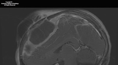
Figure 1: A mass lesion involving the skull base and pterygopalatine fossa on the left is shown on this axial FIESTA image. Abnormal soft tissue is seen in the left IAC as compared to fluid in the right IAC. The normal right greater wing of the sphenoid bone is shown. On the left, the greater wing of the sphenoid has been destroyed by the mass.
Figure 2: The soft tissue intensity skull base mass is again shown on the left. Infiltration of fat in the L pterygopalatine fossa is present, as compared with normal bright fatty signal in the R PPF. Asymmetrical signal is again seen in the left vs right IAC.
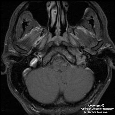
Figure 3: Following injection of gadolinium, moderate homogeneous enhancement of the mass lesion is shown. Also appreciated is extension into the L sphenoid sinus and displacement of the left cavernous segment of the L internal carotid artery. Meckel’s cave on the L is obliterated. Abnormal enhancement in the left IAC and abnormal thickening and enhancement of the tympanic segment of the facial nerve are also shown. Subtle linear enhancement extends posteriorly from the dominant mass along the greater superficial petrosal nerve on the left.
Figure 4: A more superior post-gadolinium image more discretely defines enhancement and enlargement of V3 on the left, posterior to the main bulk of the tumor mass. Tumor is again seen to extend posteriorly along the GSPN to the geniculate ganglion.
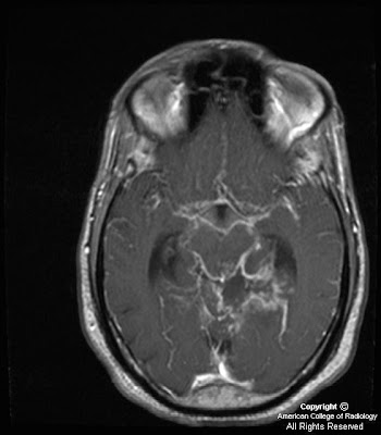
Figure 5: Tumor is seen to involve and expand Meckel’s cave on the left, replacing the normal CSF signal with intermediate signal intensity of a highly cellular tumor. The unaffected Meckel’s cave on the right, filled with CSF, is shown for comparison. The tumor is confined to Meckel’s cave on this image and has not extended through the dura to involve the adjacent temporal lobe.
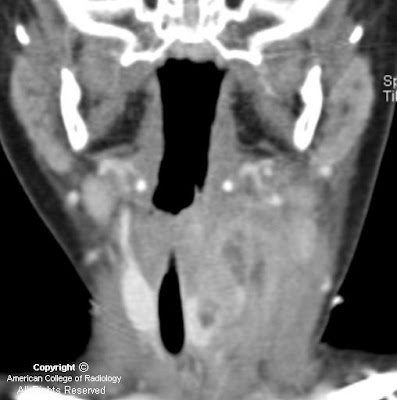
Figure 6: A more anterior coronal T2-weighted image shows extension of the tumor into the cavernous sinus on the left, adjacent to the flow void of the internal carotid artery. Marked thickening of the third or mandibular division of the trigeminal nerve is shown on the left. The mass has markedly expanded foramen ovale on the left. This image also demonstrates atrophy and mild T2 hyperintensity of the muscles of mastication on the left, due to V3 dysfunction and resultant subacute to chronic denervation change. The masticator muscles on the right have normal bulk.
Figure 7: The enhancing tumor mass involving Meckel’s cave and the cavernous sinus on the left is again shown. This image better demonstrates the unaffected foramen ovale on the right. Also demonstrated on this image is volume loss and diffuse mild enhancement of the left temporalis muscle as compared to the right, consistent with denervation change as previously discussed). Signal drop-off due to dental hardware and inhomogeneity of the magnetic field has resulted in artifact and poor fat suppression in the right masticator space.


Diagnosis: Adenoid cystic carcinoma with perineural spread of tumor


Adenoid cystic carcinomas have a high propensity for perineural invasion and extension.
The facial and trigeminal nerves are the cranial nerves most commonly involved by perineural spread of tumor.
The facial nerve may be invaded directly by tumors of the parotid gland, or the tumor may involve the trigeminal nerve and then extend along the greater superficial petrosal nerve to reach the facial nerve.
A second “five-to-seven” connection is the auriculotemporal nerve, which also provides an important route for perineural spread of tumor.
The proximal greater superficial petrosal nerve, geniculate ganglion, and tympanic segment of the facial nerve often show normal mild enhancement due to investment by a rich vascular plexus in these regions.
Characteristics of perineural extension of tumor include abnormal enhancement and enlargement of nerves, replacement of fat in neural foramina, and widening of neural foramina. Denervation changes in innervated muscles may also be observed.


Adenoid cystic carcinoma (ACC) is the second most common malignant salivary gland tumor after mucoepidermoid carcinoma. It can arise from either the major or minor salivary glands. It is the most common malignancy of the submandibular and sublingual glands. It most commonly presents as a painless enlarging mass, though the initial presentation may be due to perineural extension of tumor (pain or paresthesia, for example) if the tumor is in a deep location. ACC is associated with a high risk of distant metastases (most commonly to the lung), and these can occur 10-20 years after initial diagnosis and treatment of the primary lesion. ACC may spread through local or direct extension of the tumor, hematogenous and lymphatic dissemination, and perineural extension.

ACC has a high propensity for perineural invasion and extension. SCCs also have a high tendency to spread perineurally, and because they are the most common head and neck cancer, one will likely encounter more cases of perineural tumor spread from SCC than from ACC in practice. Other cancers of the head and neck, including melanomas, basal cell carcinomas, and mucoepidermoid carcinomas also extend perineurally, but less commonly. Branches of the facial and trigeminal nerves are most commonly involved as they innervate the cutaneous and mucosal surfaces of the head and neck, where most tumors arise, as well as the salivary glands. The facial nerve can be invaded directly when ACC occurs in the parotid gland, or the tumor may involve the trigeminal nerve and then extend along the greater superficial petrosal nerve to reach the facial nerve. An alternate route for cranial nerve 5 to cranial nerve 7 spread (or vice versa) is the auriculotemporal nerve, which is located posterior to the neck of the mandible.

The greater superficial petrosal nerve emerges from the geniculate ganglion of cranial nerve VII carrying sensory (from the soft palate mucosa) and parasympathetic fibers. It courses anteromedially through the temporal bone and emerges through the facial hiatus. It then travels underneath Meckel’s cave and combines with the deep petrosal nerve (carrying sympathetic fibers) to form the vidian nerve. The vidian nerve travels anteriorly to the pterygopalatine ganglion, where the parasympathetic fibers synapse before being distributed to the lacrimal gland and mucosal glands of the nasal and oral cavities. This nerve pathway is vulnerable to tumor infiltration and is a common pathway for perineural extension of tumor.

Portions of the facial nerve that are invested with a rich vascular plexus may normally enhance on MRI. These normally enhancing regions are the proximal greater superficial petrosal nerve, geniculate ganglion, and tympanic segment of the facial nerve; the labyrinthine and descending mastoid segments of the facial nerve may also show mild enhancement under normal circumstances. The more distal and anterior portions of the greater superficial petrosal nerve, however, are not invested by a vascular plexus and should not enhance with contrast on MRI. Similarly, the facial nerve in the IAC does not demonstrate any enhancement under normal conditions at 1.5T. Characteristics of perineural spread to the facial nerve include thickening and abnormally intense enhancement of nerve segments, as well as replacement of fat in neural foramina by tumor. Denervation changes in the muscles of facial expression may be observed, but these are often extremely subtle due the small size of the affected muscles. Due to ACC’s high propensity for perineural spread and high tendency to recur, it is important to regularly assess for perineural extension of these tumors when MR images are being interpreted.

mercoledì 20 ottobre 2010

Sialadenitis with an obstructing sialolith in the right submandibular gland duct


Findings

Coronal and axial post contrast CT images show a swollen right submandibular gland with dilatation of the intraglandular ducts and an obstructing stone.


Diagnosis: Sialadenitis with an obstructing sialolith in the right submandibular gland duct


The most common cause of sialadenitis of the SMG is an obstructing calculus with subsequent suppurative sialadenitis. Less common causes are suppurative sialadenitis leading to duct stenosis and chronic sialadenitis. Rare etiologies include include Sjogren syndrome, AIDS and bacterial/viral infection.

SMG accounts for 10% of sialadenitis of all major salivary glands. Other diagnostic considerations in SM space include reactive submandibular lymph node, mandibular osteomyelitis, benign mixed tumor, submandibular carcinoma and metastases.

Calculi are more common in the SMG duct. Compared to the parotid gland, the saliva in the SMG is thicker, much more mucinous and more alkaline. The SMG duct courses superiorly which makes it more prone to stasis. SMG duct is larger in diameter.

When sialadenitis is present therapy may depend on stone location. If the stone is in the anterior portion of the duct, the stone can be removed and gland salvaged. If stone is in the posterior duct, the duct and gland will likely be removed with the stone.

lunedì 18 ottobre 2010

Basilar meningitis of unknown etiology


Findings

Figure 1,Figure 2, Figure 3, Figure 4: Axial and sagittal T1 weighted images postcontrast demonstrate thick and nodular predominantly basilar meningeal enhancement which on sagittal images encases the middle cerebral arteries. Cerebellar parenchymal enhancement (Figure 4) secondary to late subacute infarction and infectious/inflammatory exudates.
Figure 5: Axial T2 weighted image demonstrates slight increased size of lateral ventricles with more prominent frontal horn lateral ventricles and bilateral lateral ventricle atria in a 2 week follow-up study (initial study slightly motion degraded.)


Diagnosis: Basilar meningitis of unknown etiology (presumed MDR-Tuberculosis)


This young patient with immunocompromise and altered mental status presented initially with left posterior circulation infarctions of unclear etiology. In a young patient with posterior circulation infarctions, acute vertebral artery dissection/ injury in the setting of trauma or spontaneous etiology should be considered. However, when given a history of immunocompromise and HIV, other differential diagnostic considerations such as infectious (Tuberculous, fungal, or pyogenic), inflammatory (Neurosarcoidosis), vascular (infectious vasculitis from Neurosyphilis or HIV), or neoplastic (lymphoma, leukemia, leptomeningeal carcinomatosis) etiologies should all be taken into account. To date, laboratory and CSF values for this patient continue to be non-diagnostic as to the source of this patient's basilar meningitis/vasculitis. The top consideration after discussing with the neurology team is Multi-drug resistant tuberculosis (MDR-TB) due to unimpressive response to conventional treatment regimens and inconclusive microbiological testing.

Tuberculous CNS infections are mostly caused by M. tuberculosis; atypical organisms are rare except in immunosuppressed patients. 30% of patients are HIV positive (particularly IV drug users.) Due to hematogenous dissemination, lesions usually occur at the gray-white junctions of cerebral hemispheres, basal ganglia, or cerebellum (especially in children). Hematogenous dissemination is from a systemic source, most commonly the lung, but also possibly the GU system or GI tract.

Most common manifestations of CNS Tuberculosis are meningitis, seen predominantly in the basilar cisterns, and hydrocephalus. Acutely, cerebritis can be seen, which then can progress to ring-enhancing tuberculoma lesions. Tuberculosis can also result in vasculitis and cerebral infarctions. The thick and nodular basilar meningeal enhancement in association with hydrocephalus and left posterior circulation multifocal infarctions in our patient can all be seen in tuberculosis.

Leptomeningeal sarcoidosis must be distinguished clinically from carcinomatous, lymphomatous/leukemic, and infectious meningitis. Dramatic response can be seen in some cases with steroid therapy. Sarcoid has replaced syphilis as the great mimicker. Neurosyphilis can cause arteritis of intracranial and extracranial large and medium sized arteries

giovedì 14 ottobre 2010


Findings

There is a poorly circumscribed jugular foramen mass with a permeative-sclerotic appearance of the involved bone with irregular margins and loss of the normal cortex with relative preservation of the bone density and architecture. There is centrifugal spread into the posterior fossa with a prominent dural tail and diffuse homogenous enhancement.


Jugular foramen meningioma


Jugular foramen meningiomas arise from the arachnoidal cap cells of the meninges which follow cranial nerves IX, X and XI into the jugular foramen. 5% of posterior fossa meningiomas arise in the jugular foramen and meningioma is the third most common jugular foramen mass after paraganglioma and schwannoma. If there are additional meningiomas or schwannomas, consider an association with neurofibromatosis II.

Meningiomas characteristically appear isointense to gray matter on both T1 and T2 weighted MR imaging but this appearance is variable. A relative T2 hypointensity is suggestive of a dense cellularity. There is an absence of high velocity flow voids within the mass which can help to distinguish from paraganglioma. There is dense, uniform contrast enhancement on MR and CT imaging which can help to identify a dural tail. Tumor matrix calcification is uncommon and internal hemorrhage is also rare.

Jugular foramen meningiomas typically infiltrate the surrounding skull base with relative preservation of the bone architecture. The margins of the jugular foramen are typically irregular with loss of the normal cortex. This combination of findings results in a permeative-sclerotic appearance and is different from the pemeative-destructive pattern of paragangliomas with erosion and infiltration without preservation of the underlying architecture or bone density. Additionally, schwannomas have a characteristic pattern of smooth enlargement without hyperostosis or permeative changes.

mercoledì 13 ottobre 2010

Intraorbital Lymphatic Malformation


Findings

Figure 1: There is a multilobulated mass lesion in the retroorbital region with a fluid- fluid level.
Figure 3: There is an intraconal multilobulated mass with a fluid-fluid level and mild right globe proptosis.


Diagnosis: Intraorbital Lymphatic Malformation


Vascular lesions account for 5-20% of all orbital masses, and the two most common orbital vascular lesions are venous malformations (formerly known as cavernous hemangiomas) and lymphatic malformations (LM) ( formerly known as lymphangiomas). LMs are relatively uncommon in the pediatric population and account for only 4% of all childhood orbital masses. LMs are benign and most frequently found in the head and neck. Intraorbital LMs can arise in any orbital space, but are most commonly intraconal with frequent extraconal and preseptal expansion. Histologically, a vascular malformation can contain venous and lymphatic components, hence the name lymphaticovenous malformation. Of note, 70% of orbital lymphaticovenous malformations are associated with ipsilateral, noncontiguous, intracranial vascular abnormalities.

Intraorbital venous-lymphatic malformations are present at birth, but tend not to be discovered clinically until early childhood when they enlarge as a result of either intralesional hemorrhage or lymphoid hyperplasia and result in acute proptosis. Approximately one-half of all patients with orbital LMs also complain of limited ocular mobility. Additionally, conjunctival, facial, or oral vesicles may also be observed.

Radiologic imaging of intraorbital LMs demonstrates unencapsulated, irregular, lobulated, and multicompartmental masses. These lesions can have cystic as well as more solid components. The cystic elements of these masses commonly exhibit fluid-fluid levels as a result of intralesional hemorrhage. Additionally, LMs are frequently both pre- and postseptal and intra- and extraconal. They often display orbital expansion with irregular margins that traverse tissue planes. Ultrasound images of LMs demonstrate heterogeneous, ill-defined lesions with anechoic cystic portions and extraconal extension. On CT, these masses exhibit ill-defined borders, irregular attenuations, and variable enhancement with peripheral rim enhancement in cystic regions. Additionally, calcified phleboliths can be seen on CT in venous portions of these lesions. MR imaging is the preferred imaging modality to evaluate the location, vascular components and evolving blood products of venous-lymphatic malformations. LMs demonstrate iso- to slightly high signal intensities on T1-weighted images and very high signal intensities on T2-weighted images. MR imaging also allows for the simultaneous evaluation of the brain in an effort to detect any associated intracranial vascular anomalies.

The differential diagnosis for pediatric orbital tumors can be divided into osseous and non-osseous lesions. Pediatric osseous lesions of the orbit include dermoid inclusion cysts, which are most common, fibrous dysplasia, juvenile ossifying fibroma, osteosarcoma, Langerhans cell histocytosis, granulocytic sarcoma and neuroblastoma bone metastases. Non-osseous lesions of the pediatric orbit include most commonly rhabdomyosarcoma, but additionally infantile fibromatosis, infantile hemangioma, and LM.

Orbital lymphaticovenous malformations are histologically benign, but can demonstrate aggressive behavior, such as vision loss, as they expand. The treatment of such lesions is focused on ameliorating pain, alleviating optic nerve compression, maintaining ocular alignment and improving cosmetic appearance. Surgical resection is the preferred treatment for these lesions, and while complete removal can often be achieved with well-demarcated extraconal lesions, more diffuse intraconal lesions tend to be treated with subtotal resection.

giovedì 7 ottobre 2010

Hypertrophic olivary degeneration


Findings

Figure 1 and Figure 2: Noncontrast head CT shows acute hemorrhage in the superior and middle cerebellar peduncles.
Figure 3, Figure 4, Figure 6 and Figure 7: FLAIR and T2 show chronic blood products in the superior and middle cerebellar peduncles.
Figure 5 and Figure 8: Axial FLAIR and T2 show T2 prolongation and mild enlargement
of the inferior olivary nucleus of the medulla.


Diagnosis: Hypertrophic olivary degeneration


Hypertrophic olivary degeneration is a unique type of transynaptic degeneration which results in hypertrophy rather than atrophy of the affected structure, the inferior olivary nucleus. The affected circuit involves dentrorubral-olivary connections which were described by Guillain and Mollaret in 1931 as the anatomic connections related to palatal myoclonus, and is commonly referred to as the "Guillain-Mollaret triangle."

The "triangle" consists of 3 nuclei: 1) ipsilateral inferior olivary nucleus (medulla) 2) ipsilateral red nucleus (midbrain) and 3) contralateral dentate nucleus (cerebellum). The ipsilateral red nucleus and contralateral dentate nucleus are connected by the superior cerebellar peduncle. The ipsilateral red nucleus and ipsilateral inferior olivary nucleus are connected by the central tegmental tract. There are no direct anatomic connections between the inferior olivary nucleus and the contralteral dentate nucleus.

Olivary degeneration is typically seen several months after the insult. The side of olivary degeneration depends on the location of original insult and can be predicted by familiarity with the "Guillain-Mollaret triangle." When the primary lesion is in the central tegmental tract, olivary hypertrophy is ipsilateral. When the primary lesion is in the superior cerebellar peduncle or dentate nucleus, the olivary hypertrophy is contralateral. When the primary insult involves both the central tegmental tract and superior cerebellar peduncle, the olivary hypertrophy is bilateral.

The classic clinical finding associated with hypertrophic olivary degeneration is palatal myoclonus, a cyclic jerk of the soft palate.

mercoledì 6 ottobre 2010

Labyrinthitis ossificans


Findings

Figure 1: Axial CT image through the left temporal bone shows complete ossification of the left superior semicircular canal.
Figure 3: Axial CT image at the level of the internal auditory canal shows non-erosive soft tissue attenuation lateral to the malleus and incus as well as partial ossification of the lateral semicircular canal.


Diagnosis: Labyrinthitis ossificans


Labyrinthitis ossificans (LO) is the end result of suppurative labyrinthitis where inflammation of the membranous labyrinth progresses to fibrosis and ossification. Suppurative labyrinthitis typically arises following meningitis, although other causes include direct infection from hematogenous sources or trauma.

LO is the most common cause of acquired childhood deafness and can be detected by CT as early as 2 months following an episode of meningitis. Following meningitis, approximately 6% to 30% of children develop some degree of sensorineural hearing loss which is typically bilateral.

On CT, osseous deposition is seen within the membranous labyrinth. On MRI, loss of the normal high signal on T2-weighted images from displacement of the endolymphatic fluid is suggestive of this diagnosis. It is important to the clinician to distinguish between cochlear involvement, non-cochlear involvement, or both as prognosis is determined by response to cochlear implantation. The degree of ossification is important in surgical planning and severe LO may preclude cochlear implantation.

venerdì 1 ottobre 2010

Tornwaldt cyst


Findings

There is no intracranial hemorrhage, calvarial fracture or transtentorial herniation. On the lower images of the brain, there is a cystic hypoattenuating lesion at the posterior wall of the nasopharynx measuring 1.4 x 1.2 cm.


Diagnosis: Tornwaldt cyst


A Tornwaldt cyst is a benign proteinaceous cyst that is located in the midline of the posterior nasopharynx, superficial to the superior constrictor muscle of the pharynx. It is surrounded by adenoid tissue and arises from notochordal remnants in the pharyngeal bursa (pouch of Luschka). They are seen in up to 4% of the population (equally in males and females) and are usually asymptomatic unless they become infected. If infected, they can cause a variety of symptoms including purulent drainage, sore throat, prevertebral muscle spasms, halitosis and Eustachian tube obstruction.

These fluid-filled cysts are usually discovered on imaging (both CT and MRI) as incidental findings. The cyst is well-circumscribed and located in the midline in the posterior nasopharynx (between the longus coli muscles). On CT, it is hypo-attenuating and appears cystic. It will almost invariably appear bright on T2-weighted images. The T1 signal will vary from CSF signal to very bright hyperintensity depending on the amount of protein, fat, hemorrhage and mucus within the cyst. A thin rim of peripheral enhancement may be seen with gadolinium administration. Nasopharyngoscopy, although not necessary for asymptomatic cases, can help supplement the diagnosis.

Treatment is not necessary in most cases. For the rare symptomatic cases treatment options include surgical excision, electrocoagulation or marsupialization.