

Findings
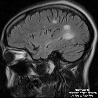
34 yo) Figure 1: Sagittal FLAIR image delineates septo-callosal interface hyperintensities, perpendicular periventricular hyperintensities extending into the deep white matter, and a juxtacortical lesion.
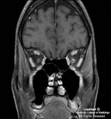
Figure 2: Coronal fat saturated T1 post gadolinium demonstrates enhancement of the left optic nerve.
38 yo)Figure 3: Sagittal FLAIR (3a) and axial FLAIR (3b) images demonstrate confluent periventricular and juxtacortical oval plaque-like hyperintense lesions perpendicular to the ventricular axis known as “Dawson’s fingers”.
44 yo) Figure 4: Axial FLAIR shows hyperintense plaques with one extending to a juxtacortical location. Figure 5: Sagittal T2 shows hyperintense plaques within the brainstem and upper cervical spinal cord. Figure 6: Sagittal FLAIR demonstrates periventricular and subcortical hyperintensities.
Diagnosis: Multiple sclerosis
Multiple Sclerosis (MS) is a demyelinating inflammatory CNS disorder of unclear etiology. It specifically affects oligodendrocytes, thus eliminating their supportive function to the neurons they serve. Women are affected twice as frequently as men, usually between the ages of 20 and 40 years. MS is a clinical diagnosis based on history, neurological examination, and paraclinical studies including MR imaging, evoked potentials and CSF studies. The hallmark of this disease is its dissemination in space and time. Diagnosis of MS separates it from clinically isolated syndromes (CIS). MS has different subtypes, the most common being the relapsing-remitting type. Other subtypes include primary progressive, progressive relapsing, and malignant/Marburg. Related demyelinating processes such as Schilder’s diffuse sclerosis and Balo’s concentric sclerosis may be considered MS subtypes. However, recurrent optic neuritis and neuromyelitis optica (Devic’s disease) have been shown to be distinctly separate entities.
Specific MR imaging characteristics include the presence of T2 hyperintensity at the septo-callosal interface and ovoid lesions perpendicular to the ventricles, known as Dawson fingers. These occur along the deep medullary veins. Active lesions may enhance avidly or poorly depending on the degree of acuity and severity. Lesions may also involve the cortex, juxtacortical white matter, brainstem, and spinal cord. These lesions, given their protean distribution and overall presentation, have an extensive differential diagnosis and therefore multiple criteria schemes have been developed to aid in the diagnosis of MS. The differential diagnosis of MS includes acute disseminated encephalomyelitis and its possible subtypes of neuromyelitis optica (Devic disease), acute optic neuritis, and acute transverse myelitis; microvascular white matter ischemic changes; progressive multifocal leukodystrophy; neurosarcoidosis; hypertensive encephalopathy; vasculitis; and encephalitis.
The 2001 International Panel on the Diagnosis of Multiple Sclerosis (IPDMS) (McDonald et al.) “McDonald Criteria” require objective evidence of CNS lesions disseminated in space and time in order to diagnose MS. Spatial criterion is defined by the Barkhof-Tintore MR imaging criteria, which require three of the following four findings: 1) at least one gadolinium-enhancing lesion or 9 T-2 hyperintense lesions; 2) at least one infratentorial lesion; 3) at least one juxtacortical lesion; 4) at least 3 periventricular lesions. Lesions should be greater than 3-mm in cross-section. A spinal cord lesion may substitute for a brain lesion, and in the setting of oligoclonal IgG bands or elevated IgG/Albumin ratio in the CSF, only 2 instead of 9 T2 lesions are needed to satisfy the criteria. Temporal criterion is satisfied by follow up imaging 3 or more months after the onset of the clinical event. The 2005 IPDMS (Polman et al.) suggested revisions to the 2001 McDonald criteria based on several research studies which followed. These modifications were made to allow for the following: 1) multiple spinal lesions may be used to substitute for brain and infratentorial lesion criteria, provided that they are more than 3mm in size, the length is less than 2 vertebral body heights, and the lesion occupies only a portion of the cord cross section, 2) an enhancing spinal cord lesion may be substituted for an enhancing brain lesion, and 3) for dissemination in time, a new T2 lesion discovery interval may be reduced from 3 months to 1 month. Polman also suggested that CSF studies are no longer needed in order to consider a diagnosis of primary progressive MS.
The 2001 IPDMS (McDonald et al.) “McDonald Criteria”
Dissemination in Space: 3 of the following 4
- At least 1 gadolinium-enhancing lesion or 9 T2-weighted hyperintense lesions*
- At least 1 infratentorial lesion
- At least 1 juxtacortical lesion
- At least 3 periventricular lesions
* + oligoclonal IgG bands or elevated IgG/Albumin ratio in the CSF, only 2 T2 lesions are needed
Dissemination in Time: One of the following
- A new enhancing lesion at least 3 months after the initial clinical event in a new clinically relevant area
- A new T2 lesion identified on a new MRI study at least 3 months after the initial scan
The 2005 IPDMS modified “McDonald Criteria”, (Polman et al.)
Dissemination in Space: 3 of the following 4
- At least 1 gadolinium-enhancing lesion (spinal cord, brainstem, and brain) or 9 T2-weighted hyperintense lesions (spinal cord, brainstem, and brain)*
- At least 1 infratentorial lesion (spinal cord, brainstem, and cerebellum)
- At least 1 juxtacortical lesion
- At least 3 periventricular lesions
* +oligoclonal IgG bands or elevated IgG/Albumin ratio in the CSF, only 2 T2 lesions are needed
Dissemination in Time: One of the following
- A new enhancing lesion at least 3 months after the initial clinical event in a new clinically relevant area
- A new T2 lesion identified on a new MRI study at least 1 month (30 days) after the initial scan
Conclusion
Diagnostic criteria for MS and its variants are continually revised as more data becomes available and MR imaging technology improves. Notwithstanding, attention to detail with precise temporospatial descriptions of lesions is essential for the diagnosis of MS and may also have prognostic value.
bueno!!
RispondiElimina